Key Requirements for A Mobile CT Provider

Key Requirements for A Mobile CT Provider

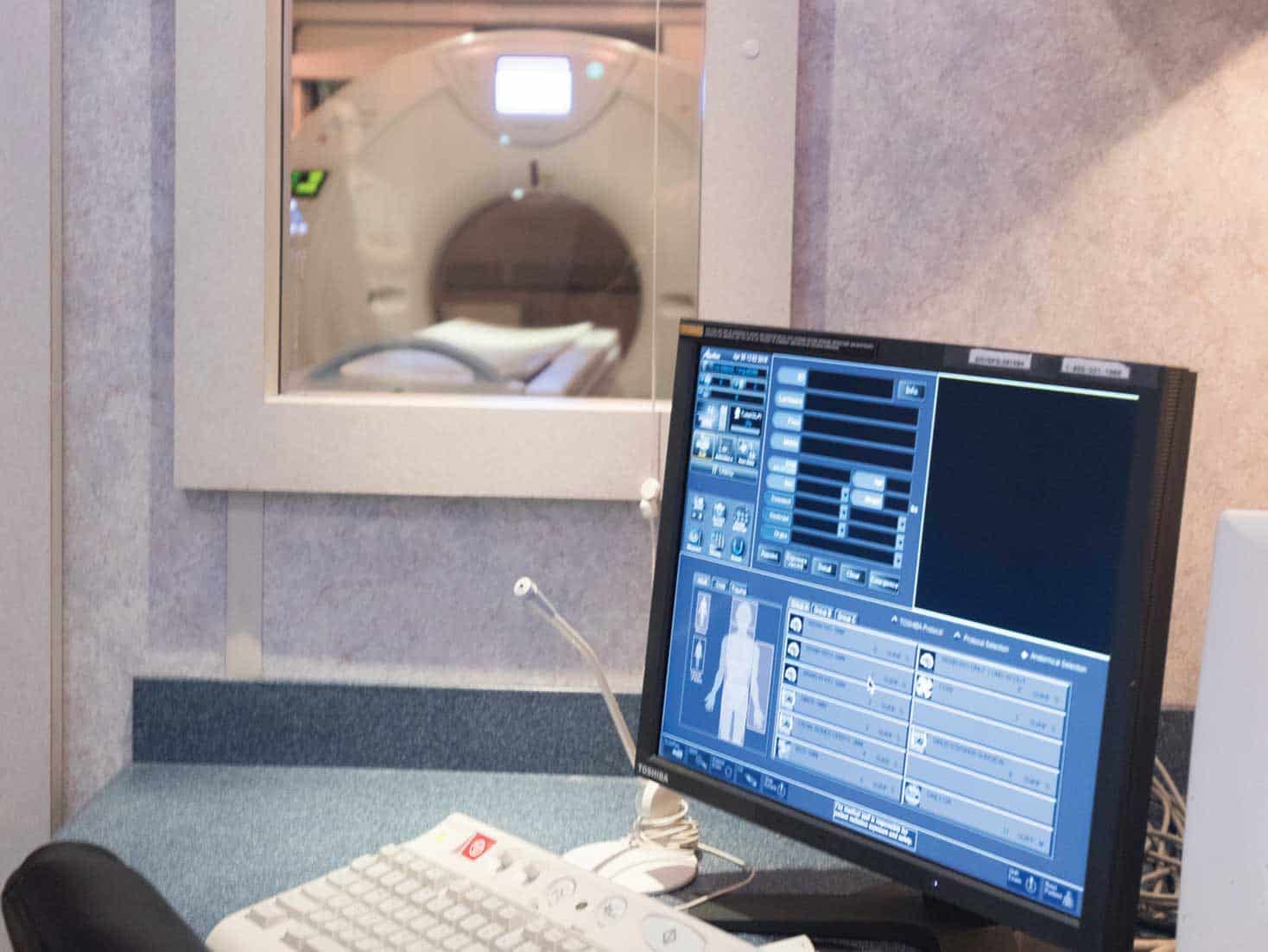
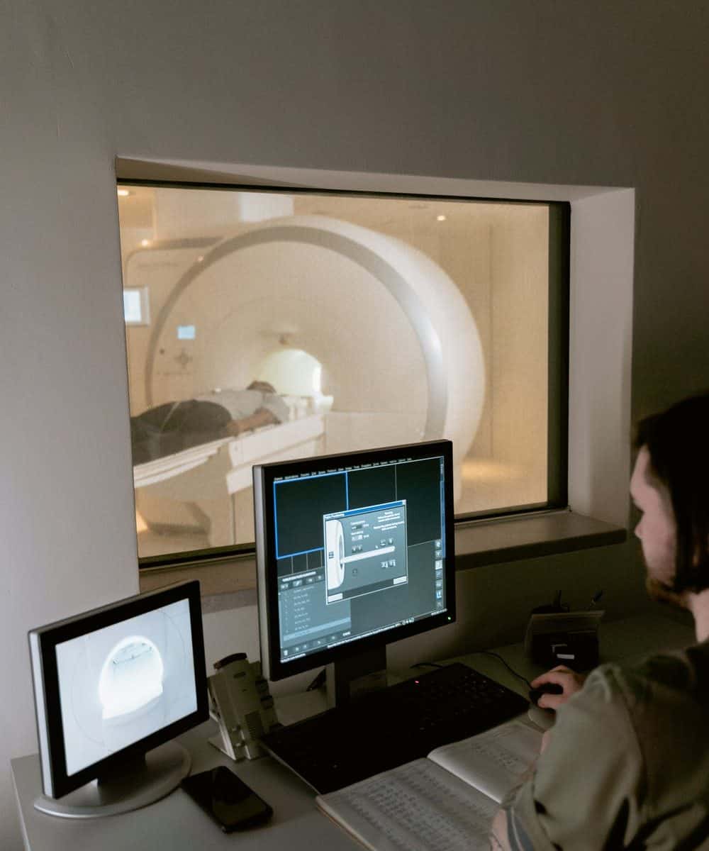
Mobile CT Requirements

Despite the fact that most hospitals have in-house CT imaging facilities, there are instances when bringing in a mobile imaging service on a temporary basis makes greater financial sense.

When redesigning your CT imaging room, when your equipment needs maintenance, or when you need assistance managing a backlog, temporary imaging might be a wise solution.

Vendors serve as an extension of your hospital’s operations.

Nonetheless, one of the most crucial considerations to bear in mind is that each vendor with whom you collaborate becomes an extension of your institution.

Your patients should not be able to tell the difference between the quality of service provided by the hospital personnel and the level of service provided by the mobile imaging team.

Maintaining the same level of cleanliness in the environment, following the same comprehensive processes, and using comparable equipment will ensure that the patient’s favorable experience continues into the mobile service setting.


What characteristics contribute to demonstrating and reinforcing a commitment to sustaining your high standards?


A list of characteristics to look for in a reputable CT mobile imaging company is provided below.

Equipment

Mobile service providers should employ equipment that is already available and frequently used in the market to meet their customers’ needs.

A 64-slice CT scanner may be preferred in certain cases, but a 16-slice camera is a perfect match for the majority of investigations and delivers outstanding picture quality while maintaining fast throughput.

Ensure that the CT scanners are XR-29 compliant as well, since non-compliance with the standard may have a negative influence on your payment.

An Original Equipment Manufacturer Service Agreement

An OEM Service Agreement should be in place for a mobile service provider in order to protect against downtime.

This assures that factory-trained service engineers are doing preventive maintenance on a regular basis and that they will have access to original equipment manufacturer (OEM) components and equipment.

Certification by the Joint Commission on Accreditation of Healthcare Organizations (JCAHO)

This independent body, known as the Joint Commission on Accreditation of Healthcare Institutions (JCAHO), is responsible for administering voluntary accreditation programs for hospitals and other healthcare organizations.

Their performance standards cover areas of the operation such as patient care, drug safety, infection control, and consumer rights, among other things.

Your mobile service provider should be JCAHO accredited, which indicates that they have implemented quality standards and practices that are above industry requirements.

HIPAA Compliance is a legal requirement.

As with your hospital’s employees, the mobile provider team should be equally dedicated to preserving patient privacy and complying with all applicable healthcare standards, including those established by the HIPAA Privacy Rule.

Check to see whether they have a HIPAA training program in place that is current and continuing.

A Sanitation and Hygiene Program

Many healthcare professionals believe that maintaining good cleanliness is the most significant technique in reducing the spread of healthcare-associated illnesses among patients and other healthcare personnel.

Make certain that the mobile service provider has rigorous hygiene rules and procedures in place, and that their employees are educated on the crucial need of infection control.


Related articles:

When is a Mobile or Modular CT Unit the Right Choice? A Definitive Guide to When You Should Do a CT Scan

Toshiba Aquillon Mobile CT Scanner Unit Station 2

There are a number of different kinds of CT scanners available. While CT scanners are often categorized as either mobile or modular, these labels don’t tell the whole story. In fact, the choice between a mobile or modular CT scanner can be quite confusing. This is because those terms are often used to describe different types of CT machines, not the specific types of scans that the machines offer.

That being said, it’s important to understand the differences between mobile and modular CT scanners in order to make the best decision for any given situation. In this article, we’re going to take a closer look at these different kinds of CT scanners, how they work, and the pros and cons of each kind of scanner.

What is a Mobile CT Scanner?

A mobile CT scanner is a device that scans tissue, either from inside the body or outside, and then processes the images so that they can be viewed on a computer monitor or sent to a device outside of the scanner. The images captured by the mobile scanners are a digital representation of the tissue being scanned, which allows for incredibly accurate and fast imagery.

The primary advantage of a mobile scanner is the amount of flexibility that it offers. They’re able to roam around where needed, without being limited by the size of any fixed area or the availability of power. They are able to scan patients who are in a variety of different positions. And, because they’re lighter and more mobile, they can be used to scan larger areas of tissue than fixed scanners.

While mobile scanners tend to be more expensive than fixed scanners, they can be less expensive than modular or active systems, if you’re willing to sacrifice some of the benefits of modularity.


What is a Modular CT Scanner?

A modular CT scanner is a CT machine that is designed to be connected to other devices to allow for new types of scanning and/or image capture. Modular CT scanners come in a number of different varieties, with most typically utilizing a variety of different sensors and cameras to scan different parts of the body.

Some of the most popular examples of modular scanners are gantry-style scanners, helical CT scanners, circular CT scanners, and helical CT scanners with integrated x-ray sources.


Mobile scanners provide a flexible solution for a variety of reasons, but one of the most important is their mobility. That mobility allows mobile scanners to often be used to scan a larger area of the body than would otherwise be possible, which can lead to better anatomical results. However, that mobility is also often coupled with higher cost, so there are trade-offs.

That higher cost is usually a result of the mobile scanner’s more advanced technology and more expensive components, as well as the more complex setup required to use it.

Modular scanners, on the other hand, offer a more cost-effective option for those who don’t need the same level of mobility that a mobile machine offers.

However, there are definitely advantages to both mobile and modular scanners.


Mobile CT Scanners

Mobile scanners are the most flexible type of CT scanner. They can be used in a variety of different ways, including outside of the body, which means that they can be used on people who are outside of a hospital setting. This is particularly important, as many different conditions require CT scans to be done outside of the body, such as on an aircraft.

Mobile scanners also offer a large field of view, which means that they can often be used to scan a very large area of tissue. This often allows for better anatomical details and a wider range of views that would be difficult to capture with a fixed scanner.

However, they also often provide higher resolution images than fixed scanners. This allows for more details and a more accurate assessment of the scan than would otherwise be possible.


Modular CT Scanners

Modular scanners are designed specifically to be connected to other devices, like computers or other electronics. They allow for a wide range of scanning options and are frequently used for special applications, like life sciences research, which requires very high-quality scans. 

Modular scanners are often specialized scanners that are designed for a specific purpose. That purpose can vary from life sciences and medical imaging, to industrial applications and more.

In fact, the first CT scanner was made for the US military, and was a modular scanner capable of scanning both inside the body and outside of it.

The most popular types of modular scanners are gantry-style scanners, which hold an x-ray source, as well as a camera or other sensor, and a computer.

Gantry-style scanners, which were originally designed for fixed hospitals, allow for a large, open workspace. This workspace can be used to scan various different parts of the body, while the source and camera/sensor can usually be moved with the scanner.


Which Mobile or Modular CT Scanner Should You Choose?

There are a number of factors to consider when choosing which kind of mobile or modular scanner to go with. Some of the most important things to consider are the size of the workspace available and the amount of mobility you need for your scans.

The mobility of a scanner impacts how large an area of tissue it can image, as well as the quality of the images that it can capture. It’s important to remember that mobile scanners, whether they’re mobile or modular, are often very expensive. So if you only need a mobile option, or you’re not willing to pay for a mobile scanner, a fixed scanner might be a better choice.


Related articles:


Bottom line

There are a number of different types of CT scanners available, so it’s important to understand the different options that are available to you. This article has provided a primer on the different types of CT scanners that are available, as well as some of the advantages and disadvantages of each type.

Ultimately, it’s important to remember that a mobile or modular scanner is a trade-off between mobility, cost, and the level of detail that can be captured.

If you need a scanner that can be used outside of a fixed location and you need good imaging quality, it might be worth purchasing a mobile scanner. If you only need a scanner for occasional scans for patients who are physically unable to move, or if you don’t have a lot of budget to play with, a fixed scanner might be a better option.

How Do I Deal With Rad Tech Workplace Fatigue?

Burned Out: Strategies to Address Rad Tech Workplace Fatigue

It’s a 7-minute sprint to the patient’s bedside, followed by 30 to 45 minutes of work, while still managing nursing staff and doctors. The rad tech then has to perform the same sprint and work routine, over and over again, for eight to 10 hours. How do rad techs keep it up for so long? It’s a job that requires focus, speed, and endurance.


Catalina Imaging - How Do I Deal With Rad Tech Workplace Fatigue?

Rad techs are expected to be some of the most reliable, steadfast workers in the medical field. They need to be able to work long shifts that require high levels of concentration and focus without showing signs of fatigue. However, that’s easier said than done. The fast-paced environment and multiple interruptions, not to mention the physical toll that the job takes on your body, often leads to workplace fatigue.


Fatigue, or a sensation of excessive tiredness that comes with poor energy and affects daily activities, is common in radiology and can affect diagnostic testing accuracy, according to the American Journal of Roentgenology (AJR). 


According to the National Physician Burnout Report published by Medscape in 2020, 46% of radiologists felt burnout, putting radiology in sixth place among specialties with the most special burnout rates.


Here are some ways to avoid burnout in the rad tech workplace.

Organize Your Priorities Everyday

Practices should allow radiologists to define daily goals and stick to them, whether it’s finding time to evaluate the most complex cases, fostering greater peer-to-peer engagement, or taking essential breaks. 


Some radiologists, for example, may feel so overwhelmed by the number of cases and pictures towering over them that they are unable to take a brief break or go out for lunch. 


Simply encouraging radiologists to schedule a half-hour break from their monitors each day to have lunch or coffee with coworkers might help them obtain a new perspective and get away from the grind.


Source: https://www.itnonline.com/article/3-tips-combat-radiologist-burnout


Recognize Your Stressors

Several stressors for technicians have been discovered via research and surveys throughout the years. Inconsistent management, ineffective communication, competing priorities, overwork, work overload, a lack of work break times, time pressures, and facilities or technology are just a few examples.


Organizations must take various steps to alleviate stress, including improved communication, a focus on adequate staffing and patient scheduling, and even personal health education (i.e., sleep habits, diet, etc.). 


Increasing the usage of staff huddles, electronic communications, and even organizing brief self-care webinars, for example, can help to give clarity and raise morale.


Source:https://www.diagnosticimaging.com/view/the-time-to-combat-radiologic-technologist-burnout-is-now

Explore Using Artificial Intelligence

Artificial intelligence and machine learning might increase the number of photos read and give you more confidence in your analysis. 


Additional workflow-improving technologies, such as collaboration portals, will go a long way toward reducing your risk of burnout and assisting you in staying organized throughout the workday.


Patient satisfaction and good results have increased in organizations that have integrated AI-guided systems and enhanced workstation technologies.


Source: https://collaborativeimaging.com/2020/02/24/how-to-reduce-burnout-among-radiologists-in-6-steps/


Apply Data-Driven Feedback to Help Your Practice Grow

Your staff is supportive of your work, and you must reciprocate. Rather than providing simple feedback, all improvement feedback should be based on cold, hard facts. 


Use data as a crucial measure of workplace success, such as the number of readings, the percentage of billing accuracies, and timeliness.


Allow the evidence to speak for itself when a problem occurs. Establish good key performance indicators for measuring your success and realistic administrative routines in your practice if you work with other health institutions. 


Determine what should be assessed in the health system other than turnaround time and agree on target metrics. Clear expectations also lower the chance of becoming overburdened.


Source: https://collaborativeimaging.com/2020/02/24/how-to-reduce-burnout-among-radiologists-in-6-steps/

Treat Yourself Weekly, Go On Vacation Trips

After a long day at the workplace, radiologists must take personal chances to relax and de-stress. 


Radiologists should avoid excessive alcohol and other harmful drugs; however, a glass of wine once in a while may be appropriate. 


Everything hinges on your capacity to develop healthy coping skills and prevent mental tiredness, which is a forerunner to burnout. Also, set aside one day every week to avoid going to the health center at all costs. 


It may not appear to be a significant difficulty, but everyone needs at least one day away from work each week.


You may also like: Why Choose Catalina Imaging

A Final Word on Avoiding Burnout In Rad Tech Workplace

“The Radiology Department has grown so much, but I’m consistently on call and feel like the department is understaffed”, said no rad tech ever. 


Radiology has a way of pulling the best out of people because it’s one of the busiest departments in the hospital. However, it does come with its own set of difficulties. It’s that constant pressure that can wear you down and make you wonder where you see yourself in the radiology department.


There are many things that are different from a normal workplace that put a rad tech at risk for workplace fatigue. With the technology and tools we have today, there are some things that are changing. It is important that we make sure we have the resources to help us cope with our work environment.


What Are The Benefits Of Using Mobile CT Imaging?

Glimpse into the Future How AI Technology Will Affect Radiology | blog articleby Catalina Imaging

Mobile CT Imaging services assist care practitioners in performing various necessary tests, actively monitoring a patient’s status, and being adaptable enough to serve patients in any setting. 


In the previous years, CT (computerized tomography) Imaging and how hospitals and health care systems use it have changed dramatically. While the market may have begun with CT scanners mounted on trucks to give services to businesses that lacked the volume to justify purchasing their equipment, it has since developed.


Today, the medical field is relying more and more on Mobile CT Imaging services to be able to deliver the best care possible to their patients. Mobile CT imaging services are making it more convenient for patients to receive the benefits of CT imaging without the need to travel to a centralized location, which can ultimately improve care and save both time and money in the long run.


What Are The Benefits Of Using Mobile CT Imaging?


The benefits of using Mobile CT Imaging in healthcare are notable. The technology is an invaluable tool that allows the specialists to save time and money. Moreover, it provides remote access to highly qualified specialists, and it is an efficient way to monitor patients.


You may also like: What Are The Trends in Computed Tomography Imaging to Look Forward to (This 2022)?


Here are just a few of the benefits of using diagnostic Mobile CT Imaging.


Faster Diagnosis

Many patients understand the aggravation of waiting for a CT scan for an extended amount of time. These delays can occur at a facility’s imaging department or at a third-party supplier that must be visited separately. 


Mobile CT Imaging is easy to scale and may be modified regularly to meet a facility’s changing needs. Rapid diagnosis is a crucial driver of mobile CT imaging. A piece of portable scanning equipment may collect images at a patient’s bedside in a fraction of the time it takes to carry a patient to a radiology room. A physician can quickly access images on the unit’s console or the PACS.


Beyond severely ill patients in the ICU, providing diagnoses faster and more efficiently benefits a significant portion of the patient population. Patients’ care with life-threatening injuries, for example, can be scanned immediately at their bedside rather than risking transport problems. 

Lower Cost of Health Care

An ounce of prevention is better than a pound of cure. Doctors can fix problems once they’ve been properly identified and diagnosed. Nuclear medicine diagnostics, for example, can detect cardiac abnormalities before they progress to the point where surgery is required. 


Late-stage diseases and disorders frequently need more rigorous therapy or surgery, resulting in higher costs and additional risks. However, when conservative treatment options are used early on, your timely intervention will likely reduce your healthcare expenditures.


Improved Patient Treatment 

Transferring severely sick patients to the radiology department might result in further difficulties. According to the International Journal of Critical Illness & Injury Science, transport complications can include possible danger to the respiratory system, hemodynamics (including loss of intravenous access), traumatic injury, acid-base homeostasis, glucose regulation, infection risk, and even death.


Bringing diagnostic CT imaging technology to the patient’s bedside can avoid complications from transporting a patient. It also removes the need for transportation personnel, allowing them to focus on other patients in the hospital.


The contribution of mobile CT imaging to patient care is the key reason why many healthcare institutions are adopting it today. 

Painless Diagnostic Procedures

Mobile CT Imaging is a relatively new standard in diagnostic procedures. It is used to scan the interior of the human body, providing doctors with a non-invasive way to detect possible health issues. 


This reduces the need for invasive procedures, the most important being the complete lack of radiation. The technology is an invaluable tool that allows specialists to save time and money. 


Moreover, it provides remote imaging that can be used in cases of emergencies, for example, when the patient is unable to physically visit the doctor.


It’s important to note that “mobile” is not just a word that describes the ability to be moved around. Instead, it is an expression of the fact that the CT scanner can be used in a wide range of situations. This means that doctors don’t have to worry about patients trying to get to the clinic. Instead, they can use the scanner to examine the patient in the most convenient location. This could mean the patient’s home, a hospital or in a remote area.

Infection Prevention & Decrease of Radio Exposure

Infection control is always a problem in the hospital setting, but it is more so now with the arrival of COVID-19. Monitoring patients at their bedside can help prevent contagions from spreading during patient transfer.


Imaging devices are known to emit radiation. In an effort to reduce the number of x-rays taken in a hospital and to prevent the spread of infections, many hospitals are turning to Mobile CT Scanning. 


While out-of-hospital CT scanning is not new, it is gaining more popularity as hospitals try to minimize the number of x-rays they take per patient while still providing a high level of patient care. As a result, many hospitals have invested in mobile CT scanners.


For large facilities, the radiation dose can be significant when CT is being used on multiple patients. A Mobile CT Scanner can help reduce doses by allowing for a dedicated scanner that can be used for multiple patients. This can help reduce the overall exposure of patients to radiation and also help decrease the need for transporting patients and their potentially infectious body products to other locations. 


You may also like:


A Final Word on the Benefits of Mobile CT Imaging 

When it comes to health care, the number one goal is always patient safety. With this in mind, Mobile CT Imaging makes it possible for hospitals and doctors to deliver higher quality care to their patients. 


The most important part about Mobile CT Imaging is that it helps doctors diagnose and treat their patients more quickly. This means that if a patient is having a serious medical emergency, they can receive the care they need faster. The results are better patient outcomes and fewer unnecessary tests.


Catalina Imaging is committed to providing the highest quality solutions by specializing in Mobile CT Scanners using state-of-the-art Siemens, GE, and Toshiba/Canon technology. Give our team a call today at (844) 949-1664.

What Are The Trends in Computed Tomography Imaging to Look Forward to (This 2022)?

What Are The Trends in Computed Tomography Imaging to Look Forward to (This 2022)?

The computed tomography (CT) scanning procedure is a type of specialized imaging that helps physicians view organs and bones non-invasively. CT scanners route X-rays through the body, capturing multiple angles of a cross-sectional image to compile into a 3D representation which can be stored and examined on computers. The data is processed using algorithms to reconstruct images in the human-readable format.


Catalina Imaging - Featured Images


Function and productivity will drive many of the trends we’re seeing for CT imaging this 2022. As the sophisticated technology behind computed tomography continues to spread its reach, users can now enjoy greater access to these tools through third party companies specializing in their manufacture and distribution. 


For example, Mordor Intelligence forecasts that by 2026, the CT market will grow by 5.76 percent annually, hitting a hefty $9.5 million by that time.


While rates of cancer, including those for colorectal and cervical cancer, are thankfully dropping in the United States and other developed countries, cancer remains a persistent health crisis. In response to the increased demand observed for several years now for CT scans that diagnose many different cancers, scanner manufacturers have begun enhancing their flagship models with more productivity-enhancing features.


Some of the recent trends in CT Imaging technology seen this past year include:


Quality, Dose and Workflow

Philips announced the release of Spectral CT 7500, which has received regulatory clearance from both Europe and the U.S. FDA. This system promises to deliver high-quality spectral images 100 percent of the time without needing special protocols like those on many other newest models, subsequently reducing protocol bias and confirming old information in up to 60 percent fewer tests than ever before. 


The Spectral CT 7500’s  intelligent software eliminates all of the guesswork behind acquiring test scans, giving radiologists more accurate data faster than they’ve ever had before. 


Philips’s goal with Spectral CT 7500 is to help physicians better characterize disease and reduce rescans, allowing them to detect abnormalities earlier on with just as much precision as conventional CT scans but with less radiation exposure for patients overall.


Artificial Intelligence Technologies

GE Healthcare released its Revolution Ascend with Effortless Workflow, which offers clinicians a collection of AI technologies that automate and simplify time-consuming tasks to increase operational efficiency and free up time for clinicians to deliver more personalized care for more patients. 


Equipped with a lower table, 40mm detector coverage, and a larger, new 75 cm wide-gantry, the Revolution Ascend is designed to accommodate high body mass index (BMI) patients, as well as trauma cases that would otherwise be too delicate to maneuver in a smaller size gantry.


Other features built into GE’s Revolution Ascend:

  • GE’s Effortless Workflow, a new suite of AI solutions that personalizes scans accurately and automatically for each patient, and requires significantly less effort from the CT technologist. 
  • Auto Positioning, which uses real-time depth-sensing technology to generate a 3-D model of the patient’s body and uses a deep learning algorithm to determine the correct table elevation and cradle movements to align the center of the scan range with the isocenter of the bore.
  • The Clarity Operator Environment, a suite of embedded intelligent tools that can provide optimal scan range settings, dose and image quality for each patient, helping to deliver greater efficiency and more personalized medicine across clinical care areas.


Intelligent User Interface

The FDA recently approved an innovative single-source CT scanner by Siemens Healthineers called the Somatom X.ceed. Its scanner is designed for all scanning procedures and is aimed at providing state-of-the-art diagnostic imaging through an 82 cm bore.


The Siemens Healthineers Somatom X.ceed features both high-speed scanning capabilities and a level of resolution previously unseen in other single-source CT systems, with a new hardware/software combination to simplify CT-guided interventions. 


Key features of the Siemens Healthineers Somatom X.ceed:

  • a fast rotation speed of 0.25 seconds to ensure a high native temporal resolution and reduce motion artifacts when scanning, moving structures such as the heart. 
  • a scan speed of 262 mm/sec to provide consistent image quality across the entire field of view
  • a small focal point on the scanner–  0.4 x 0.5– to enable increased spatial resolution to better detect deep-seated small and medium lesions. 
  • a 1,300 mA power reserve, for higher power, enabling a high level of image quality for larger patients while expanding the utilization of low dose and low contrast media techniques, such as low kV imaging.
  • myNeedle Companion combines hardware innovations and intuitive software to coordinate the planning and guidance of percutaneous needle procedures. 
  • myExam Companion user guidance software employs artificial intelligence to help simplify and standardize scanner operation for each user. It also provides a new decision tool for trauma imaging. 
  • myExam Satellite feature allows immediate assessment of CT perfusion results, which can be very valuable in emergency radiology.


Accuracy, Precision and Speed

The new Aquilion Exceed LB CT system from Canon Medical Systems USA Inc. has just received FDA clearance, The Aquilion’s  CT technology delivers high-quality, high-resolution volumetric, 3D reconstruction images in a clinically relevant and cost-effective manner. 


The Aquilion Exceed LB CT system has the potential to enhance oncologists’ treatment of cancer patients because of its ability to reduce radiation dose exposure, increase patient throughput and improve accuracy. 


The system was specifically designed for the radiotherapy market to provide better visualization of tumor anatomy during treatment planning with uniform patient positioning that enables accurate target localization for all types of cancer.


Features of the Aquilion Exceed LB CT system include: 

  • Canon Medical’s Advanced intelligent Clear-IQ Engine (AiCE) Deep Learning Reconstruction (DLR) technology, which provides sharper, clearer, and more distinct images. 
  • a large bore opening (90 cm),
  • edge-to-edge extended field-of-view (90 cm) reconstruction 
  • and wide detector coverage (4 cm). 


You may also like:


A Final Word on CT Imaging Trends This 2022

Function and productivity will drive many of the trends we’re seeing for CT imaging this 2022. 


One of the things that will continue to drive the industry forward will be the need for faster exam times. The faster exams can help reduce patient stress, improve physician efficiency, and increase a facility’s throughput. 


We will likely see more manufacturers continue to implement and improve on existing workflow enhancements. Workflow enhancements can include faster gantry rotation, faster table speed, and faster reconstruction times to name a few. These enhancements are all focused on helping to drive exam times down. 


We will also see the adoption of more advanced software and hardware applications such as CAD, virtual reality, and automated patient positioning. These applications will help to improve the workflow and patient experience, but also help to improve the final image quality.


Source: Imaging Technology News 

Healthcare… Why are we in this business?

Doctor with patient using CT scan

What is it that drives someone to want to be in healthcare? Is it the humanity, knowing you are contributing to a family having more knowledge in their loved one’s health issues? Could it be the money? Markets show an industry increase of over 800 billion for 2021. With so much growth, this must mean you can make a lot of money, right?


Whether your reasons are purely altruistic or not, we all share one thing in common. WE DO IT!


For me, healthcare has been a passion of mine since I was 18, when my father passed away from pancreatic cancer. During his treatment, I remember the feeling of complete helplessness. I can recall thinking to myself, what can I do to make a difference. It took some years, but I found my spot in healthcare.


My first venture into healthcare was as a Field Service Engineer (FSE), repairing medical equipment all over the US. My new venture provides a solution to hospitals. This enables hospitals to maintain the same level of care while their CT is being repaired or replaced. In some cases, it offers a hospital an inexpensive alternative to building a new hospital suite. At the end of the day, I know I am doing what I was intended to do, help patients obtain answers.


Misconceptions in CT Imaging

Patient Awareness Drives CT Scan Needs | Catalina Imaging

With over  40 years of clinical experience in diagnostic imaging and the last 18 as a vendor CT Clinical Application Specialist, I have noticed some common misconceptions among CT Technologist which I would like to discuss. Misconceptions, which if not understood can lead to higher than necessary patient exposure as well as a degradation in CT image quality. There are also misconceptions about terminology which I hope to clear up as each vendor has their own specific terminology for processes in CT which mean the same thing, such as scout, pilot and topogram just to make a simple one.


It doesn’t matter how a patient lies down on the CT couch.

Patients should be carefully positioned at gantry isocenter. So many times while conducting CT application training, I have observed technologist not carefully or properly positioning patients on the CT couch to isocenter within the gantry. This is so imperative for a couple reasons. Number one being the x-ray exposure to the patient and number two being image quality.

The X-ray exposure to the patient is calculated from the AP and Lateral scout views, along with many other scanning and reconstruction factors, determined by the system software. The patient needs to be positioned at gantry isocenter because this is where the computer will read the density of the anatomy being imaged (notice I did not say patient size!). Tube Current modulation in modern CT systems offer offer a modulation scheme which includes adjustment of the mA in an X and Y direction or angular, as well as Z axis or longitudinal mA modulation. This type of mA modulation adjusts the exposure to the size and shape of the patient’s anatomy, which will result in the proper exposure for the requested image quality setting within the imaging protocol, the noise value. If the patient is too high in the scan field of view or too low in the scan field of view, the exposure software reading the scout images will make an incorrect assessment of anatomical densities. Too high in the scan field of view will lean to a higher CT exam exposure to the patient. Conversely, to low in the scan field of view will lead to a CT exposure which is too low to maintain the image quality setting within the scan parameters. Scan field of view choices should be selected based on the patient’s body habitats and the patient carefully positioned within the scan field to gantry isocenter. If the patient is not correctly positioned in the scan field of view within the system gantry, both X-ray exposure and image quality will be affected.


This is also because of the bow-tie filter incorporated in the system, which is chosen automatically with the scan field of view. The purpose of a bow-tie filter is to attenuate the X-rays at the elliptical skin surface of the patient and thus reducing exposure to those areas. A patient positioned in an off center lateral or vertical direction will lose the advantages of the bow-tie filter.

The source to image receptor distance in CT systems is a constant which does not change. The X-ray source to gantry isocenter is also a constant. The variable is with patient positioning either being too high relative to gantry isocenter or too low. An increase of the object to detector distance will result in a proportional increase in magnification of the radiographic image. Since in CT, data is collected for image reconstruction from 360 degrees of angulation around the patient, the patient must be properly positioned to avoid image distortion within the reconstruction process.


Image reconstruction has no effect on patient exposure.

Modern CT system have some very intuitive exposure control systems which take into account not only the size, positioning and density of the patient being scanned, they also look at noise levels within an image data set, reconstruction algorithms and reference or reconstruction slice widths.

A noise level or standard deviation, is set in the scan parameters which exposure control systems use to calculate how much the mA can modulate through the scan range while maintaining the specific noise level or standard deviation within the reconstructed data set for the reference slice width. This sets the signal to noise ratio for the scan. Reference slice width or a reconstruction slice width also plays a part because thinner slice widths are inherently noisier than thicker slice widths and therefore would require the system to put out more energy (signal) to maintain the set noise level in a thinner reconstructed slice width.

Reconstruction algorithms also effect patient exposure when using automated exposure controls because some reconstruction algorithms are much more edge enhancing than others. Reconstructions such as lung or bone to use extreme examples. Other subtle differences in reconstruction algorithms may be experienced by using the reconstruction algorithms of a particular vendors system. For the same body region, say soft tissue for example, you may see different algorithm choices. Some more sharp some more smooth, some with more or less contrast and some with larger or smaller grain size depending on the likes of the reading radiologist. Depending on your system, these reconstruction choices may be built within an exposure control default setting, or on an exposure control tab or an image reconstruction tab which the technologist sets prior to scanning the patient. This varies by vendor and vendors software advances which automates the exposure control systems.

All CT vendors will provide application support to your facility with the purchase of a new system. As the system is being used by many technologist over its years of use it may have been modified away from the manufacturers default settings, thus negatively affecting patient exposure and image quality. Scheduling an applications visit from a qualified specialists should take place periodically and is just as important as routine system maintenance.

There are other considerations when employing a mobile CT solution at a facility. Mobile CT systems are moved from facility to facility, and the scanning and reconstruction parameters set on the system could have been set by some well-meaning but very uninformed technologist. As the user of a mobile system there should be some insistence that the service engineer setting up the mobile unit reinitialize the system defaults and scanning protocols so you are starting off with an imaging system scanning as designed by the manufacturer. Not doing so, is to rely on the staff using the system prior to arriving at your facility to have set it up correctly, which most likely, (and in my experience) will not be the case. Using a system which is not optimized to the manufacturers specifications can lead a facility to false impressions regarding a given vendor or equipment. Radiologist may complain regarding image quality or have issues with patient exposure, so it’s always a good idea to have the system properly set up. If you are using a mobile CT solution on a temporary basis while a new system is being installed at your facility, it would be beneficial to your staff to have a system from the same vendor as the system being installed. This will assist the staff with learning the new system operating software, should you be switching CT vendors. This gives your staff a leg up on the transition. Have the mobile company supply a few days of application support to assure your staff technologist and physicians are happy with the system operation and to train staff with building scanning protocols, raw data reconstruction, MPR and any other post processing necessary for your facility. It is important that the person doing the training is very qualified, and familiar with the selected mobile system.


Automatic generation of coronal and sagittal data sets.

Positioning the patient properly will give an improvement of image quality with symmetry to the axial plane, and the correct automatic generation of coronal and sagittal views.

Modern CT scanners have the ability to improve technologist workflow by allowing for the automatic generation of coronal and sagittal views within a scanning protocol. These views however are actually generated in a reconstruction slice width and interval from the top of the display field of view to the bottom of the display field of view and we call these images coronal views when the patient is lying supine on the CT couch. The same is true of the sagittal image data set. It is re-constructed from right to left or left to right depending on how the protocol is determined. However, these views are only true direct coronal or true direct sagittal image data sets when the patient is properly positioned on the CT couch and properly aligned to the laser positioning lights. So many times patients are positioned and scanned on the couch in a slight oblique and/or rotated plane. This leads to sub optimal image data sets being sent to a radiologist who then must make a diagnosis for the patient. Since it takes only a few minutes to make sure that the patient is properly positioned and aligned to the laser positioning lights, all CT technologist should not avoid this important preliminary step. Proper patient positioning is necessary for assuring the right exposure to the patient, and the best image quality for the radiologist. It is important to remember that a technologist should always ask the patient for permission to touch them and explain why prior to moving the patient to align them with the positioning lasers.


Higher kVp, is it better for CT Angiography, CTA?

When performing CT angiographic studies, so often I come across scan protocols utilizing a very high kVp of 130-140. I have heard technologist say that it because you have to penetrate the contrast, or that the body part is thick and dense. In my experience, patients with a BMI of 30 or less, 100 kVp will penetrate the patient’s anatomy and will yield to a more contrasting image data set because the kVp is closer to the K edge of the iodine contrast. A technologist will see an increase in the mA value set by the systems exposure control system as it compensates to maintain the signal to noise ratio set in the protocols. In my experience that little bump in mA, which yields more photons to the detector, will produce a lower CTDI than which would be achieved using 120-130-140 kVp.

Since we are discussing CT angiograms, helical pitch is another scan parameter which is misunderstood in CT angiography. Modern CT systems usually have table speed options for helical pitch which include a slow table speed which would provide for a smaller available scanning range but with higher image detail and image quality and dose to the patient, to a fast table speed giving a longer scan range with a shorter total scan time. A slow table speed is not desirable for CT angiograms because you could lose your contrast bolus, increase venous contamination in the image set and leads to a higher than necessary patient exposure. Use either a standard pitch or a faster pitch, and remember your exposure control system will compensate with a bump in the mA value. For example, I like a faster table speed and 100 kVp for a PE study because I can capture the beating heart with less motion artifact in the lung fields and in the ascending aorta. The lower kVp means that the IV contrast on my images will be brighter because it is less penetrated.


How does Scan Field of View affect neuro image quality?

The CT image we view on the computer screen is made up of CT Pixels displayed over the CT matrix. It is the field of view divided by the matrix which determines the size of the pixels in the displayed image. Keeping in mind that the smaller the pixel size, the greater the special resolution, CT technologist should endeavor to scan brain/neuro studies in the smallest field of view that will encompass the patient’s anatomy, in this case the head. This usually requires more precisely positioning the patient’s head at gantry isocenter. So many times I have witnessed staff technologist move the scan range either more anterior, posterior or laterally to encompass the patient’s anatomy because it was not properly positioned within the scan field which can be quite small, 240mm in some cases. This moving of the scan acquisition results in the system shifting the scan parameters from a small scan field of view, say 240 mm to a slightly larger field of view (320-400mm) which changes the pixel size. Scanning in a larger field of view will create larger pixels thus compromising spacial resolution required for brain and neuro imaging. Technologist should keep in mind that it is better to re-position the patient within the small scan field of view and not continue the habit of moving the scan field of view to the patient’s position. This is also necessary for proper CT exposure reporting. Brain scans should have a dose calculation based on a 16cm water phantom and there is a possibility that a change in the scan field of view could change the exposure calculation. When scanning multiple anatomical areas using the same set of scout images, such as a head and neck or C spine, the technologist should carefully center to the patient’s head as that has the smallest field of view requirement for both proper spatial resolution and exposure reporting.


Thin overlapping image reconstruction is not Raw Data.

Thin overlapping images have a wide variety of uses from the creation of simple coronal and sagittal data sets without stair stepping artifacts caused by thick data sets, to MIP and MINIP data sets created automatically on the CT system. Thin overlapping data sets are loaded into sophisticated 3D workstations and advanced diagnostic computer applications such as Cardiac, Perfusion and Dual Energy. These thin image data sets, (since they are reconstructed images), are at times wrongly referred to as Raw Data. Rule of thumb, if you can see it, it is image data. This misrepresentation may be because the thin data set is often reconstructed at the raw data or acquired slice width, however with an overlap by as much as 50%. This type of reconstruction improves Z axis resolution while limiting partial volume averaging. Since these data sets are reconstructed images, they are not Raw Data.

The term Raw Data should be reserved only to describe the digital data collected from the scan acquisition prior to its reconstruction process. This Raw Data is held by the system and is made available to the operator for further manipulation until such time that it is automatically overwritten or the operator deletes it, if it is not protected.

Raw Data is used and manipulated by the operator when there is required a change in the display field of view, reconstruction algorithms, to change the start and end point of a reconstruction, a change of slice width, interval or any change in X and or Y component of a reconstructed image data set.


MPR is not a “reconstruction”.

MPR is often referred to as a Multi Planer Reconstruction, as many technologists use this term to refer to the coronal, sagittal, oblique or curved images made from the thin overlapping axial image data sets. We should consider that MPR is actually a Reformat (“to give a new format to, revise or represent in another format”. Definition from Oxford Language Dictionary), of the thin axial images to a change of projection to the coronal, sagittal, oblique or curved plane. The first use of Reformat according to Merriam Webster was in 1967 as a transitive verb meaning: “To format (something) again or in a different way.” Since image data is used and manipulated (reformatted in a different way) to a different plane of view, Reformat or Reformation would be the proper term for this kind of image.

What to Look for in a CT Scanner Service Provider

If you are a radiologist manager or hospital administrator, choosing the “right” mobile CT scanner service provider might be a daunting task because you have to consider the service fee, the type of equipment they offer, and the nitty-gritty details of their contract.

To help you choose the right service provider, we compiled a list of things you should always consider.

Experience

When we say experience, we are not just referring to the number of years a CT scan engineer has spent to hone his expertise, but also the service provider’s experience in handling a specific model and brand. For this reason, a GE CT engineer with a decade of knowledge may not be the best one to work on a Siemens scanner (and vice versa).

The general rule of thumb is to ask the provider’s “preferred” CT scan brands and models if you want to assess their level of expertise and experience.

Availability of parts

It is advisable to choose a service provider that stocks their own inventory of mobile CT scanner parts. In this way, they won’t have a hard time finding a replacement part in the event that this medical equipment breaks down.

Providers that keep their own replacement parts can help you save money because you don’t have to pay for the shipping cost. Additionally, you don’t have to wait for days, or worse, weeks, just for the parts to arrive on your doorstep.

Simply put, you can avoid additional expenses and prolonged downtime if you partner with a service provider that keeps their own replacement part inventory.

Customer Service

Always choose a provider that offers a customer-focused service, meaning they should maintain an open line of communication, including weekends and holidays. In addition, they should be able to resolve problems (for example, the CT scan needs repair or tune-up) as quickly as possible. Hence, you may want to partner with a local service provider whose engineers live just a short drive from your facility.

Reasonable Service Fee

Opt for a CT scan service provider that offers reasonable service fees and discounts. For instance, some companies offer a discount if you agree to a long-term lease, i.e., a minimum of one year.

Of course, price should not be the only consideration when choosing a provider. Keep in mind that the quality of service trumps all other factors.

Links:

To learn more about high-quality CT scanner providers, call Catalina Imaging at (844) 949-1664 or schedule your free consultation by clicking here.

Catalina Imaging‘s commitment is to provide your facility with industry-leading mobile solutions to ensure your scanning needs are met. We offer you state-of-the-art technology supported by our dedicated customer service, guaranteeing efficiency when it is needed most.

Reasons Why Medical Facilities Rent Mobile CT Scanners

Toshiba Prime 160-slice Mobile CT Scanner

Most hospitals and medical facilities that choose to rent mobile CT scanners than purchase this medical imaging equipment do so because of the upfront cost. As of this writing, the cost of new ct scanners greatly varies between $285,000 and $2.1 million.

Why Do Medical Facilities Rent Mobile CT Scanners?

They are running a training or education course.

Hospitals and medical facilities that offer training often need to rent a mobile CT scanner, so their own imaging equipment is not tied up to their education course. Usually, they opt for a long-term lease (a minimum of one year) to enjoy a huge discount.

They need a trial run.

Newly established medical facilities may want to conduct a “trial run” before they purchase their own [fixed] CT scan, which of course, involves a considerable investment that can reach millions of dollars. Once they can establish sustainable foot traffic–i.e., enough patients who need this imaging procedure–they may proceed to buy their own scanner.

On the other hand, if the trial run shows that the number of patients every month fluctuates significantly, they may want to stick to the leasing arrangement at least for the meantime.

Their fixed CT scanners are under renovation or maintenance.

If the hospital’s current CT suite is under maintenance or renovation, they need to rent this mobile CT scan to continue providing this imaging procedure that is commonly used in detecting injuries, abnormal growth, and damage in bones, blood vessels, and internal organs.

There is a backlog of patients.

Some medical facilities experience a sudden spike of backlogs that affect the quality of patient care. Hence, hospitals rent CT scanners from time to time to make sure that they can keep up with the demand.

Purchasing a fixed CT scanner may not be a sound investment.

Some hospital networks prefer to rent mobile CT scanners than to equip each site with its own CT suite to save money. This is ideal if not every medical facility site receives enough foot traffic to justify buying an expensive imaging equipment.

When partnering with hospital networks, a mobile CT rental can have its trailer run a route and then stop at each site for a couple of days.

They need to preserve their working capital and cash flow.

Having a fixed CT suite is a huge investment that can reach millions of dollars. For this reason, small hospitals and medical facilities opt for a long-term lease to preserve their working capital and cash flow.

Leasing laboratory equipment is usually ideal for medical facilities that need to reinvest more capital into the core operations of their business.

(Note: Medical machinery is generally hard to resell, which further makes it ideal to lease it than purchase one if the medical facility needs to improve their working capital.)

They want to improve patient care.

In the past, people from small towns needed to travel long distances just to undergo CT scans from city hospitals. But with the advent of mobile CT scanners, patients will simply need to go to their local hospitals to receive this imaging procedure.

Related articles:

To learn more about our mobile CT scan rental, call us at (844) 949-1664 or visit Catalina Imaging to know the benefits of partnering with us.

Links:

Patient Awareness Drives CT Scan Needs

Patient Awareness Drives CT Scan Needs | Catalina Imaging

Patient Awareness of Different Imaging Test Options 

Over the recent years, around 75 million CT scans have been performed annually in the US, a number that is expected to continue to reach 84 million by 2022, according to iData Research. 


Patient awareness is one of the key drivers of the growing number of CT scan procedures and other non-invasive medical imaging technologies. This is particularly true for individuals with a strong [family] history of cancer and other debilitating diseases who can benefit from early detection and diagnosis, which in turn increases the success rate of their treatments. 


However, doctors would not perform CT scans and other medical procedures without a substantial medical benefit or if these are not medically indicated. 


Patients should bear in mind that the physicians’ job is not only to diagnose a disease and perform treatments but also to determine whether a test is medically warranted in the first place. 


CT Provider: What Patients Look For 

With the advent of the Internet, today’s patients are more aware of their options when it comes to healthcare and treatment. Furthermore, they demand a patient-centric experience that includes non-invasive procedures that provide quick and accurate results. 


Patients are also aware of the radiation risk that comes with CT scans, prompting them to choose providers that adjust the settings of their scanners based on their body size and age. For children, it is particularly important to limit their radiation exposure because of their small frame and increased sensitivity to radiation. 


Modern CT scanners can provide optimal image quality even at lower radiation levels, says leading mobile CT provider Catalina Imaging


To further promote high patient satisfaction, Catalina Imaging encourages good communications between patients and their healthcare providers, adding that patient experience also plays a significant role in adherence to treatment and its effectiveness and outcomes. 


GE Healthcare also echoes Catalina Imaging’s stance on empowering patients and improving communications to provide them with a better experience. 


On its website, GE Healthcare states that technologists should perform these guidelines to improve patient experience: 

  • Acknowledge the patients and introduce themselves before the procedure. 
  • Describe the procedure and how long it takes. 
  • Provide patients opportunities to ask questions. 
  • Express genuine concern and empathy toward their patients. 
  • Acknowledge patient fear and anxieties. 
  • Communicate to the patients that they can be reached during a procedure. 


Better Imaging Technology

Today’s CT scanners can scan through large areas of the body in just a few seconds, even with lower levels of radiation and contrast dye. Such speed has been tied to improved patient experience, according to several surveys. 


Another notable advancement in medical imaging procedures is the use of mobile CT scanners, which are housed within a trailer that allows them to move from one medical facility to the next.


Mobile CT scanners have made quality healthcare more accessible to patients in rural areas who were initially forced to travel long distances to go to city hospitals. 


Catalina Imaging: The Leading Mobile CT Scanner Provider 

Catalina Imaging has been at the forefront of the campaign for a better patient experience during medical imaging procedures. Through their nationwide fleet of mobile CT scanners, they have been helping healthcare facilities make medical imaging tests more accessible to residents near them. 


Their service and storage locations are in Loomis CA, Upland CA, Forest Lake MN, Mokena IL, and Statesville NC


For more information, you can call them at (844) 949-1664 or send an email at info@catalinaimaging.