How Do I Deal With Rad Tech Workplace Fatigue?

Burned Out: Strategies to Address Rad Tech Workplace Fatigue

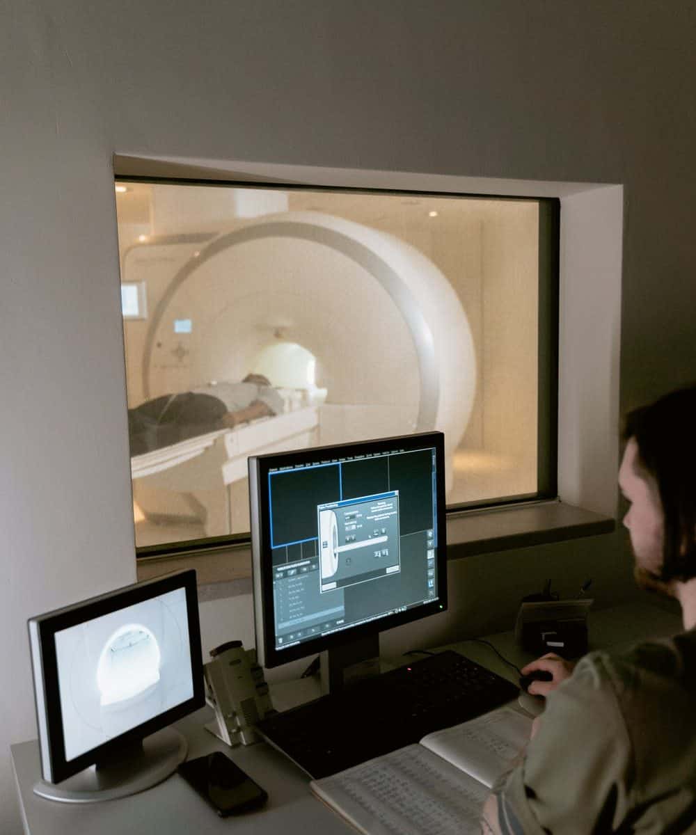
It’s a 7-minute sprint to the patient’s bedside, followed by 30 to 45 minutes of work, while still managing nursing staff and doctors. The rad tech then has to perform the same sprint and work routine, over and over again, for eight to 10 hours. How do rad techs keep it up for so long? It’s a job that requires focus, speed, and endurance.


Catalina Imaging - How Do I Deal With Rad Tech Workplace Fatigue?

Rad techs are expected to be some of the most reliable, steadfast workers in the medical field. They need to be able to work long shifts that require high levels of concentration and focus without showing signs of fatigue. However, that’s easier said than done. The fast-paced environment and multiple interruptions, not to mention the physical toll that the job takes on your body, often leads to workplace fatigue.


Fatigue, or a sensation of excessive tiredness that comes with poor energy and affects daily activities, is common in radiology and can affect diagnostic testing accuracy, according to the American Journal of Roentgenology (AJR). 


According to the National Physician Burnout Report published by Medscape in 2020, 46% of radiologists felt burnout, putting radiology in sixth place among specialties with the most special burnout rates.


Here are some ways to avoid burnout in the rad tech workplace.

Organize Your Priorities Everyday

Practices should allow radiologists to define daily goals and stick to them, whether it’s finding time to evaluate the most complex cases, fostering greater peer-to-peer engagement, or taking essential breaks. 


Some radiologists, for example, may feel so overwhelmed by the number of cases and pictures towering over them that they are unable to take a brief break or go out for lunch. 


Simply encouraging radiologists to schedule a half-hour break from their monitors each day to have lunch or coffee with coworkers might help them obtain a new perspective and get away from the grind.


Source: https://www.itnonline.com/article/3-tips-combat-radiologist-burnout


Recognize Your Stressors

Several stressors for technicians have been discovered via research and surveys throughout the years. Inconsistent management, ineffective communication, competing priorities, overwork, work overload, a lack of work break times, time pressures, and facilities or technology are just a few examples.


Organizations must take various steps to alleviate stress, including improved communication, a focus on adequate staffing and patient scheduling, and even personal health education (i.e., sleep habits, diet, etc.). 


Increasing the usage of staff huddles, electronic communications, and even organizing brief self-care webinars, for example, can help to give clarity and raise morale.


Source:https://www.diagnosticimaging.com/view/the-time-to-combat-radiologic-technologist-burnout-is-now

Explore Using Artificial Intelligence

Artificial intelligence and machine learning might increase the number of photos read and give you more confidence in your analysis. 


Additional workflow-improving technologies, such as collaboration portals, will go a long way toward reducing your risk of burnout and assisting you in staying organized throughout the workday.


Patient satisfaction and good results have increased in organizations that have integrated AI-guided systems and enhanced workstation technologies.


Source: https://collaborativeimaging.com/2020/02/24/how-to-reduce-burnout-among-radiologists-in-6-steps/


Apply Data-Driven Feedback to Help Your Practice Grow

Your staff is supportive of your work, and you must reciprocate. Rather than providing simple feedback, all improvement feedback should be based on cold, hard facts. 


Use data as a crucial measure of workplace success, such as the number of readings, the percentage of billing accuracies, and timeliness.


Allow the evidence to speak for itself when a problem occurs. Establish good key performance indicators for measuring your success and realistic administrative routines in your practice if you work with other health institutions. 


Determine what should be assessed in the health system other than turnaround time and agree on target metrics. Clear expectations also lower the chance of becoming overburdened.


Source: https://collaborativeimaging.com/2020/02/24/how-to-reduce-burnout-among-radiologists-in-6-steps/

Treat Yourself Weekly, Go On Vacation Trips

After a long day at the workplace, radiologists must take personal chances to relax and de-stress. 


Radiologists should avoid excessive alcohol and other harmful drugs; however, a glass of wine once in a while may be appropriate. 


Everything hinges on your capacity to develop healthy coping skills and prevent mental tiredness, which is a forerunner to burnout. Also, set aside one day every week to avoid going to the health center at all costs. 


It may not appear to be a significant difficulty, but everyone needs at least one day away from work each week.


You may also like: Why Choose Catalina Imaging

A Final Word on Avoiding Burnout In Rad Tech Workplace

“The Radiology Department has grown so much, but I’m consistently on call and feel like the department is understaffed”, said no rad tech ever. 


Radiology has a way of pulling the best out of people because it’s one of the busiest departments in the hospital. However, it does come with its own set of difficulties. It’s that constant pressure that can wear you down and make you wonder where you see yourself in the radiology department.


There are many things that are different from a normal workplace that put a rad tech at risk for workplace fatigue. With the technology and tools we have today, there are some things that are changing. It is important that we make sure we have the resources to help us cope with our work environment.


What Are The Benefits Of Using Mobile CT Imaging?

Glimpse into the Future How AI Technology Will Affect Radiology | blog articleby Catalina Imaging

Mobile CT Imaging services assist care practitioners in performing various necessary tests, actively monitoring a patient’s status, and being adaptable enough to serve patients in any setting. 


In the previous years, CT (computerized tomography) Imaging and how hospitals and health care systems use it have changed dramatically. While the market may have begun with CT scanners mounted on trucks to give services to businesses that lacked the volume to justify purchasing their equipment, it has since developed.


Today, the medical field is relying more and more on Mobile CT Imaging services to be able to deliver the best care possible to their patients. Mobile CT imaging services are making it more convenient for patients to receive the benefits of CT imaging without the need to travel to a centralized location, which can ultimately improve care and save both time and money in the long run.


What Are The Benefits Of Using Mobile CT Imaging?


The benefits of using Mobile CT Imaging in healthcare are notable. The technology is an invaluable tool that allows the specialists to save time and money. Moreover, it provides remote access to highly qualified specialists, and it is an efficient way to monitor patients.


You may also like: What Are The Trends in Computed Tomography Imaging to Look Forward to (This 2022)?


Here are just a few of the benefits of using diagnostic Mobile CT Imaging.


Faster Diagnosis

Many patients understand the aggravation of waiting for a CT scan for an extended amount of time. These delays can occur at a facility’s imaging department or at a third-party supplier that must be visited separately. 


Mobile CT Imaging is easy to scale and may be modified regularly to meet a facility’s changing needs. Rapid diagnosis is a crucial driver of mobile CT imaging. A piece of portable scanning equipment may collect images at a patient’s bedside in a fraction of the time it takes to carry a patient to a radiology room. A physician can quickly access images on the unit’s console or the PACS.


Beyond severely ill patients in the ICU, providing diagnoses faster and more efficiently benefits a significant portion of the patient population. Patients’ care with life-threatening injuries, for example, can be scanned immediately at their bedside rather than risking transport problems. 

Lower Cost of Health Care

An ounce of prevention is better than a pound of cure. Doctors can fix problems once they’ve been properly identified and diagnosed. Nuclear medicine diagnostics, for example, can detect cardiac abnormalities before they progress to the point where surgery is required. 


Late-stage diseases and disorders frequently need more rigorous therapy or surgery, resulting in higher costs and additional risks. However, when conservative treatment options are used early on, your timely intervention will likely reduce your healthcare expenditures.


Improved Patient Treatment 

Transferring severely sick patients to the radiology department might result in further difficulties. According to the International Journal of Critical Illness & Injury Science, transport complications can include possible danger to the respiratory system, hemodynamics (including loss of intravenous access), traumatic injury, acid-base homeostasis, glucose regulation, infection risk, and even death.


Bringing diagnostic CT imaging technology to the patient’s bedside can avoid complications from transporting a patient. It also removes the need for transportation personnel, allowing them to focus on other patients in the hospital.


The contribution of mobile CT imaging to patient care is the key reason why many healthcare institutions are adopting it today. 

Painless Diagnostic Procedures

Mobile CT Imaging is a relatively new standard in diagnostic procedures. It is used to scan the interior of the human body, providing doctors with a non-invasive way to detect possible health issues. 


This reduces the need for invasive procedures, the most important being the complete lack of radiation. The technology is an invaluable tool that allows specialists to save time and money. 


Moreover, it provides remote imaging that can be used in cases of emergencies, for example, when the patient is unable to physically visit the doctor.


It’s important to note that “mobile” is not just a word that describes the ability to be moved around. Instead, it is an expression of the fact that the CT scanner can be used in a wide range of situations. This means that doctors don’t have to worry about patients trying to get to the clinic. Instead, they can use the scanner to examine the patient in the most convenient location. This could mean the patient’s home, a hospital or in a remote area.

Infection Prevention & Decrease of Radio Exposure

Infection control is always a problem in the hospital setting, but it is more so now with the arrival of COVID-19. Monitoring patients at their bedside can help prevent contagions from spreading during patient transfer.


Imaging devices are known to emit radiation. In an effort to reduce the number of x-rays taken in a hospital and to prevent the spread of infections, many hospitals are turning to Mobile CT Scanning. 


While out-of-hospital CT scanning is not new, it is gaining more popularity as hospitals try to minimize the number of x-rays they take per patient while still providing a high level of patient care. As a result, many hospitals have invested in mobile CT scanners.


For large facilities, the radiation dose can be significant when CT is being used on multiple patients. A Mobile CT Scanner can help reduce doses by allowing for a dedicated scanner that can be used for multiple patients. This can help reduce the overall exposure of patients to radiation and also help decrease the need for transporting patients and their potentially infectious body products to other locations. 


You may also like:


A Final Word on the Benefits of Mobile CT Imaging 

When it comes to health care, the number one goal is always patient safety. With this in mind, Mobile CT Imaging makes it possible for hospitals and doctors to deliver higher quality care to their patients. 


The most important part about Mobile CT Imaging is that it helps doctors diagnose and treat their patients more quickly. This means that if a patient is having a serious medical emergency, they can receive the care they need faster. The results are better patient outcomes and fewer unnecessary tests.


Catalina Imaging is committed to providing the highest quality solutions by specializing in Mobile CT Scanners using state-of-the-art Siemens, GE, and Toshiba/Canon technology. Give our team a call today at (844) 949-1664.

What Are The Trends in Computed Tomography Imaging to Look Forward to (This 2022)?

What Are The Trends in Computed Tomography Imaging to Look Forward to (This 2022)?

The computed tomography (CT) scanning procedure is a type of specialized imaging that helps physicians view organs and bones non-invasively. CT scanners route X-rays through the body, capturing multiple angles of a cross-sectional image to compile into a 3D representation which can be stored and examined on computers. The data is processed using algorithms to reconstruct images in the human-readable format.


Catalina Imaging - Featured Images


Function and productivity will drive many of the trends we’re seeing for CT imaging this 2022. As the sophisticated technology behind computed tomography continues to spread its reach, users can now enjoy greater access to these tools through third party companies specializing in their manufacture and distribution. 


For example, Mordor Intelligence forecasts that by 2026, the CT market will grow by 5.76 percent annually, hitting a hefty $9.5 million by that time.


While rates of cancer, including those for colorectal and cervical cancer, are thankfully dropping in the United States and other developed countries, cancer remains a persistent health crisis. In response to the increased demand observed for several years now for CT scans that diagnose many different cancers, scanner manufacturers have begun enhancing their flagship models with more productivity-enhancing features.


Some of the recent trends in CT Imaging technology seen this past year include:


Quality, Dose and Workflow

Philips announced the release of Spectral CT 7500, which has received regulatory clearance from both Europe and the U.S. FDA. This system promises to deliver high-quality spectral images 100 percent of the time without needing special protocols like those on many other newest models, subsequently reducing protocol bias and confirming old information in up to 60 percent fewer tests than ever before. 


The Spectral CT 7500’s  intelligent software eliminates all of the guesswork behind acquiring test scans, giving radiologists more accurate data faster than they’ve ever had before. 


Philips’s goal with Spectral CT 7500 is to help physicians better characterize disease and reduce rescans, allowing them to detect abnormalities earlier on with just as much precision as conventional CT scans but with less radiation exposure for patients overall.


Artificial Intelligence Technologies

GE Healthcare released its Revolution Ascend with Effortless Workflow, which offers clinicians a collection of AI technologies that automate and simplify time-consuming tasks to increase operational efficiency and free up time for clinicians to deliver more personalized care for more patients. 


Equipped with a lower table, 40mm detector coverage, and a larger, new 75 cm wide-gantry, the Revolution Ascend is designed to accommodate high body mass index (BMI) patients, as well as trauma cases that would otherwise be too delicate to maneuver in a smaller size gantry.


Other features built into GE’s Revolution Ascend:

  • GE’s Effortless Workflow, a new suite of AI solutions that personalizes scans accurately and automatically for each patient, and requires significantly less effort from the CT technologist. 
  • Auto Positioning, which uses real-time depth-sensing technology to generate a 3-D model of the patient’s body and uses a deep learning algorithm to determine the correct table elevation and cradle movements to align the center of the scan range with the isocenter of the bore.
  • The Clarity Operator Environment, a suite of embedded intelligent tools that can provide optimal scan range settings, dose and image quality for each patient, helping to deliver greater efficiency and more personalized medicine across clinical care areas.


Intelligent User Interface

The FDA recently approved an innovative single-source CT scanner by Siemens Healthineers called the Somatom X.ceed. Its scanner is designed for all scanning procedures and is aimed at providing state-of-the-art diagnostic imaging through an 82 cm bore.


The Siemens Healthineers Somatom X.ceed features both high-speed scanning capabilities and a level of resolution previously unseen in other single-source CT systems, with a new hardware/software combination to simplify CT-guided interventions. 


Key features of the Siemens Healthineers Somatom X.ceed:

  • a fast rotation speed of 0.25 seconds to ensure a high native temporal resolution and reduce motion artifacts when scanning, moving structures such as the heart. 
  • a scan speed of 262 mm/sec to provide consistent image quality across the entire field of view
  • a small focal point on the scanner–  0.4 x 0.5– to enable increased spatial resolution to better detect deep-seated small and medium lesions. 
  • a 1,300 mA power reserve, for higher power, enabling a high level of image quality for larger patients while expanding the utilization of low dose and low contrast media techniques, such as low kV imaging.
  • myNeedle Companion combines hardware innovations and intuitive software to coordinate the planning and guidance of percutaneous needle procedures. 
  • myExam Companion user guidance software employs artificial intelligence to help simplify and standardize scanner operation for each user. It also provides a new decision tool for trauma imaging. 
  • myExam Satellite feature allows immediate assessment of CT perfusion results, which can be very valuable in emergency radiology.


Accuracy, Precision and Speed

The new Aquilion Exceed LB CT system from Canon Medical Systems USA Inc. has just received FDA clearance, The Aquilion’s  CT technology delivers high-quality, high-resolution volumetric, 3D reconstruction images in a clinically relevant and cost-effective manner. 


The Aquilion Exceed LB CT system has the potential to enhance oncologists’ treatment of cancer patients because of its ability to reduce radiation dose exposure, increase patient throughput and improve accuracy. 


The system was specifically designed for the radiotherapy market to provide better visualization of tumor anatomy during treatment planning with uniform patient positioning that enables accurate target localization for all types of cancer.


Features of the Aquilion Exceed LB CT system include: 

  • Canon Medical’s Advanced intelligent Clear-IQ Engine (AiCE) Deep Learning Reconstruction (DLR) technology, which provides sharper, clearer, and more distinct images. 
  • a large bore opening (90 cm),
  • edge-to-edge extended field-of-view (90 cm) reconstruction 
  • and wide detector coverage (4 cm). 


You may also like:


A Final Word on CT Imaging Trends This 2022

Function and productivity will drive many of the trends we’re seeing for CT imaging this 2022. 


One of the things that will continue to drive the industry forward will be the need for faster exam times. The faster exams can help reduce patient stress, improve physician efficiency, and increase a facility’s throughput. 


We will likely see more manufacturers continue to implement and improve on existing workflow enhancements. Workflow enhancements can include faster gantry rotation, faster table speed, and faster reconstruction times to name a few. These enhancements are all focused on helping to drive exam times down. 


We will also see the adoption of more advanced software and hardware applications such as CAD, virtual reality, and automated patient positioning. These applications will help to improve the workflow and patient experience, but also help to improve the final image quality.


Source: Imaging Technology News