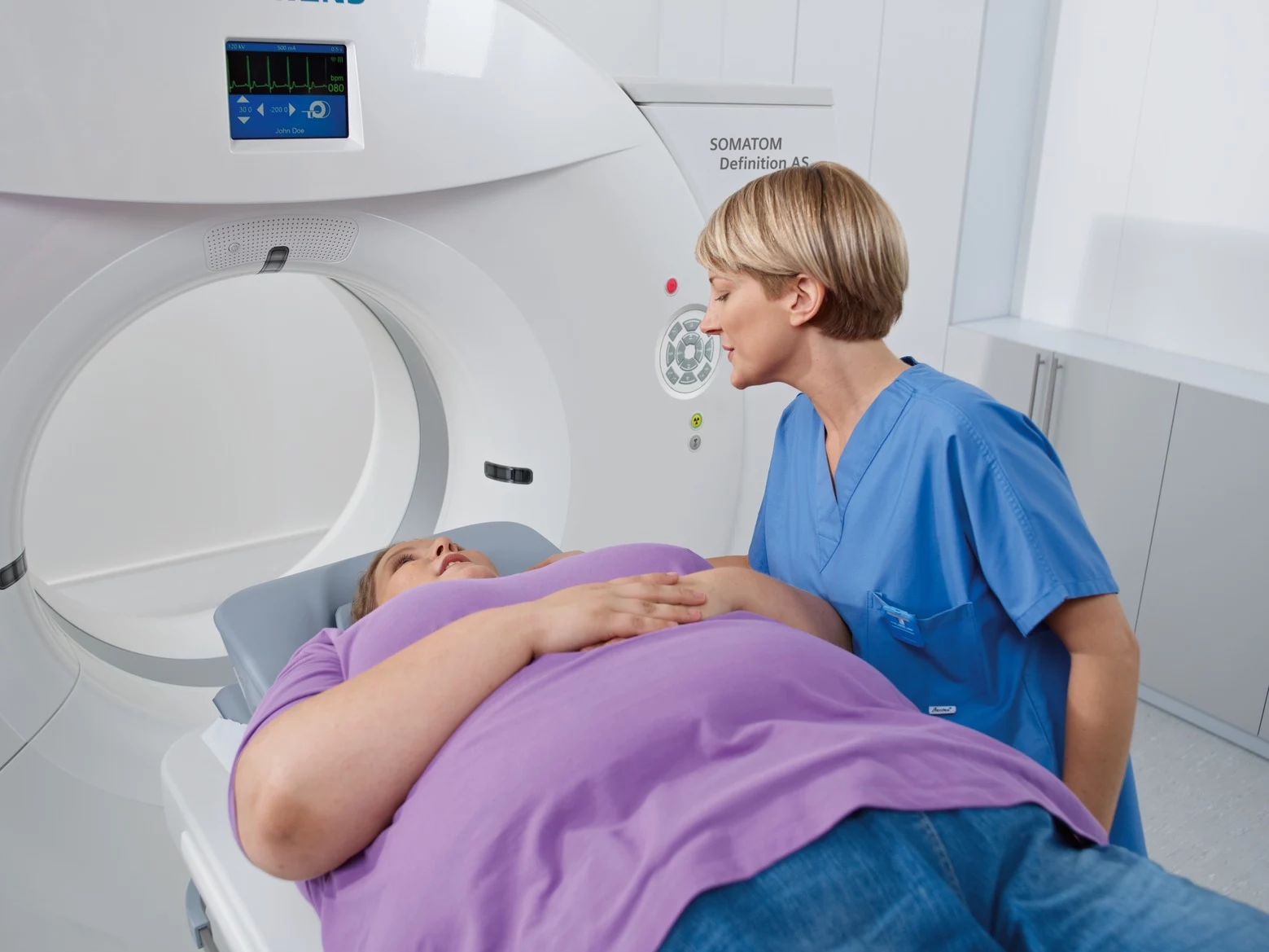
Enhancing Patient Experience: The Comfort and Design of Catalina Imaging’s Mobile Units
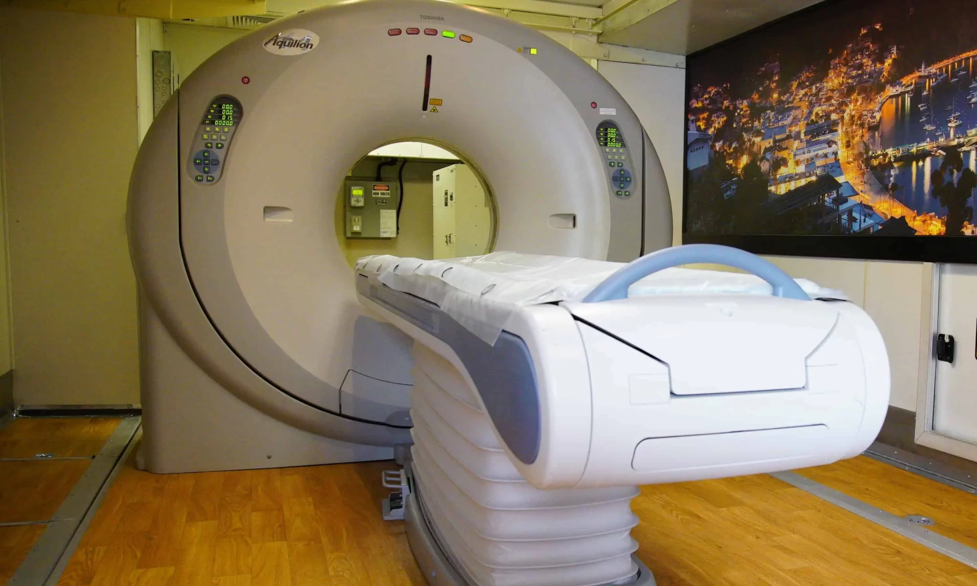
When it comes to diagnostic imaging, technology isn’t the only thing that matters – patient experience is equally vital. A well-designed imaging environment can ease anxiety, improve scan accuracy, and contribute to overall care satisfaction. That’s why Catalina Imaging goes beyond technical performance when delivering its mobile CT scanners.. They...
Continue reading "Enhancing Patient Experience: The Comfort and Design of Catalina Imaging’s Mobile Units"