The Role of Mobile CT Scanners in Transcatheter Aortic Valve Replacement

The Role of Mobile CT Scanners in Transcatheter Aortic Valve Replacement | Catalina Imaging

A transcatheter aortic valve replacement (TAVR) is a life-saving, minimally invasive procedure that treats aortic stenosis, a type of heart valve disease. TAVR replaces a narrowed aortic valve, usually in cases of aortic valve stenosis where said valve fails to open properly.

The replacement valve may be made of human or animal tissue. Most importantly, the procedure does not require open-heart surgery and the entire procedure typically only takes a couple of hours.

What Happens In Cases of Aortic Stenosis?

A quick review: the main role of the human heart is to make sure the necessary amount of blood reaches various parts of the body so that it can continue performing optimally.

To help circulate the blood and direct blood flow, there are valves in our heart which are the pulmonary valve, tricuspid valve, aortic valve, and mitral valve. They all have different functions, but just like any other machine, some parts of it may break down or need to have maintenance or have some of it replaced.

Of these four, the aortic valve is most subject to narrowing given its thin tissue composition. Once this narrowing, also called stenosis, starts, the aortic valve begins to have difficulty opening to its full extent.

This narrowing causes the heart to double its effort in pumping enough blood to the body, which eventually weakens the heart muscle due to overwork.

Once aortic stenosis turns critical, the patient might start to exhibit chest pain, difficulty in breathing, and possibly fainting. It is important to get medical assistance as soon as one of these symptoms show to avoid life-threatening consequences later.

What Causes The Aortic Valve To Narrow?

Aortic stenosis occurs when the heart’s aortic valve thickens and calcifies. The valve does not open fully and blood flow from the heart to the rest of the body is limited.

If you experience fainting, chest pain, irregular heart rhythm, fatigue, and shortness of breath, you may have aortic valve stenosis.

This is what happens when the valve narrows and the flow of blood are diminished.

whole aorta CT scan taken during a transcatheter aortic valve replacement (TAVR) procedure using a GE Revolution HD unit.

Who Might Need to Undergo Transcatheter Aortic Valve Replacement?

Aortic stenosis is a common diagnosis among the elderly, affecting over two million people in the US alone. Most cases are caused by calcium buildup in the valve due to old age, and these patients do not show symptoms until they reach 70 or above.

In some instances, however, people can develop aortic stenosis as a complication from congenital heart defects or rheumatic fever. If left untreated, this condition can lead to infections in the heart, blood clots, strokes, and ultimately, heart failure.

Getting regular check-ups will significantly help to monitor the valves’ condition, and early detection will also positively affect the results of treatment.

Treating Aortic Stenosis

It was in 2002 that the first TAVR procedure was carried out successfully and it continuously developed thanks to technology, technique, and experience.
Before, general anesthesia was used in Europe between 2002 and 2008, but after a few studies, local anesthesia with conscious sedation was the default in TAVR surgeries.

Also, initially, surgical cut down was used to carry out the procedure, but over the past few years, percutaneous closure was adopted because it was less invasive and posed lower risks. In addition to this, CT scan or angiography is also used now to check the common femoral artery site before the procedure, and during the TAVR surgery, while the puncture is being done.

The treatment for aortic stenosis depends on the severity of the case.

For patients showing mild stenosis, there is no required treatment yet and they can still proceed normally with their daily activities. Since aortic stenosis is a progressive condition, they will still need to undergo regular monitoring to make sure that if their case gets worse, they can receive due and proper care.

Meanwhile, patients with moderate stenosis should refrain from undergoing strenuous physical actions like running, weightlifting, and core-heavy exercises. They will also need to take annual medical evaluations, as further medication might be necessary depending on the status of the aortic valve.

For instance, there are cases wherein patients will be given antibiotics if valve infection is found.

When aortic stenosis reaches severe status, the most effective way of treatment is to completely replace the affected valve.

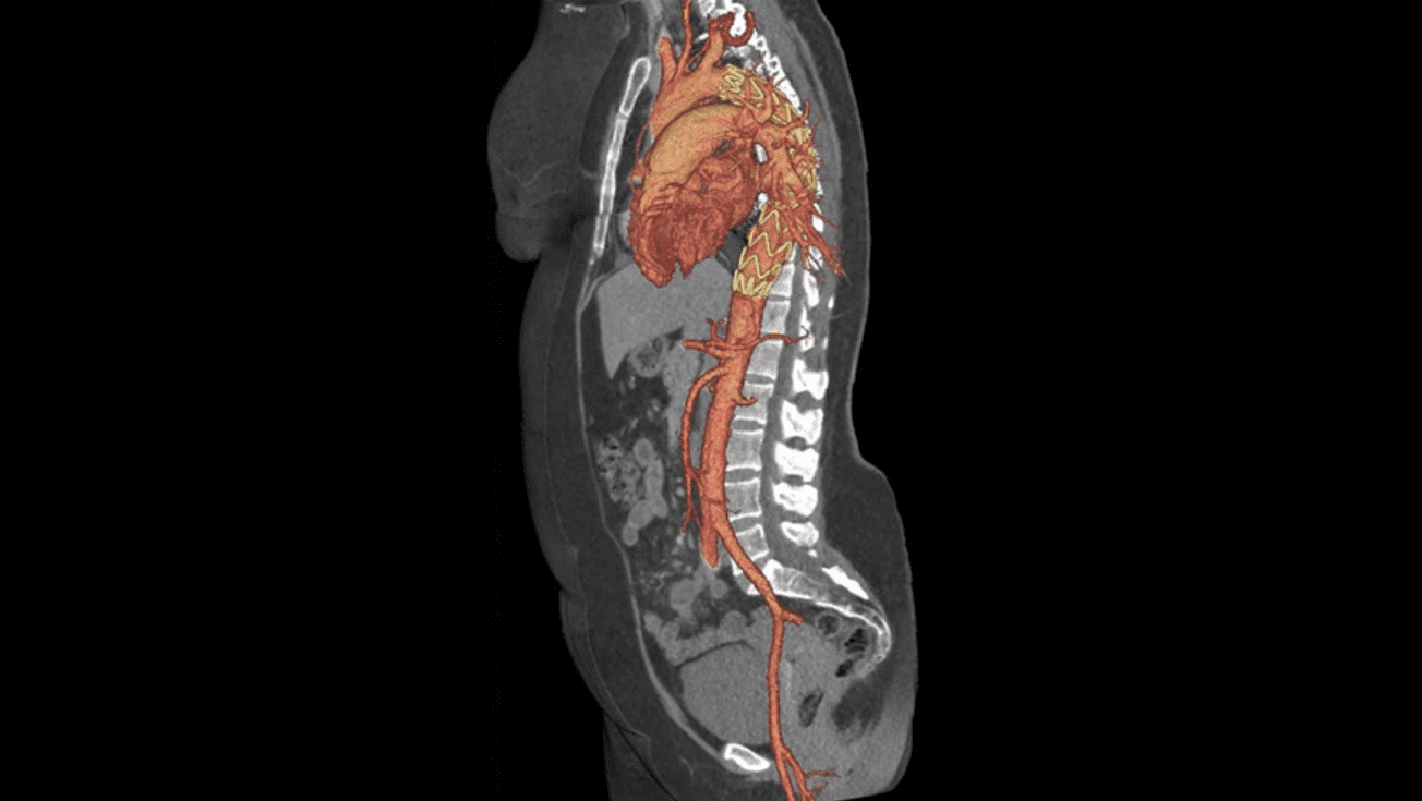
CT Scanners and Transcatheter Aortic Valve Replacement

There were limited options for valve replacement before. Most of these operations were done using open-heart surgery, wherein doctors will make a surgical opening in the middle of the chest to gain access to the heart. They will then take out the narrowed valve and replace it with a new one.

However, John Hopkins Medicine claims that this procedure can be dangerous for people with advanced age or underlying conditions like diabetes, kidney or lung disease, previous heart operations, or stroke history. In such cases, Transcatheter Aortic Valve Replacement (TAVR) might be better suited for the patient.

TAVR provides beneficial treatment options to people who may not have been considered for valve replacement. The procedure is available to patients in all risk categories.

However, might be too risky for patients to undergo the procedure if they have the following conditions:

  • Advanced age
  • Chronic obstructive lung disease
  • Diabetes
  • Kidney disease
  • Large calcium deposits in blood vessel
  • Previous heart surgery
  • Radiation treatment to the chest
  • Stroke history
  • Weaker heart

whole aorta CT scan taken during a transcatheter aortic valve replacement (TAVR) procedure using a GE Revolution HD unit.

Preparing for the TAVR Procedure

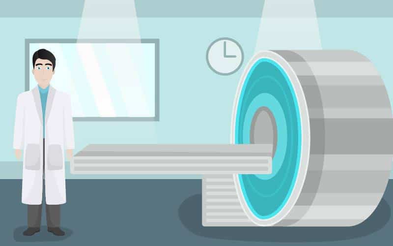
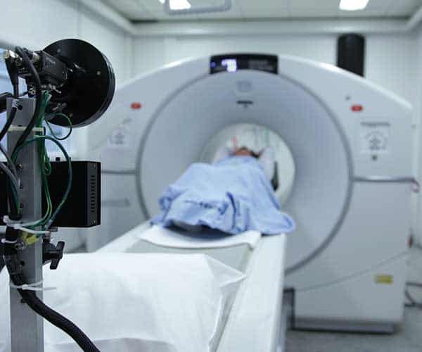
In TAVR, there is less risk because the doctors will not have to open the chest at all. Prior to surgery, the medical team will use a computed tomography scan (a CT scan) to create an image of the heart.

Recommendations for the sizing and reporting of the aortic valve, annulus, and outflow tract are diagnosed by the CT scanner, as well as reporting of fluoroscopic angulation, vascular access, coronary artery, and even non-cardiac, non-vascular findings. These are essential in determining what is needed during TAVR.

These scans will help them evaluate the heart’s condition and map out a way to the valve.

As with any other major surgery, one will need to be prepared for transcatheter aortic valve replacement. You will need to do the following:

  • Full medical check (x-rays, CT scans, blood tests)
  • Echocardiogram
  • Cardiac catheterization

For the food and drug restrictions:

  • All the drugs you’re currently taking should be communicated to your healthcare provider
  • If you’re a smoker, you will need to stop a few days before your surgery, so mention this to your healthcare provider
  • The midnight before your surgery, stop eating and drinking

The Transcatheter Aortic Valve Replacement Procedure

In a TAVR procedure, a new valve is inserted without removing the old, damaged valve. The new valve is placed inside the diseased valve.
Usually, valve replacement requires an open-heart procedure with a “sternotomy”, in which the chest is surgically separated (opened) for the procedure. This isn’t the case anymore with TAVR, which is favorable for more patients.

During operation, a small incision will be made, normally in the groin area, for the doctor to insert a catheter– a thin, flexible tube– into the femoral artery (a transfemoral approach). Sometimes, the doctor may opt to enter via a small incision in the chest and entering through a large artery in the chest or through the tip of the left ventricle (the apex), which is known as the transapical approach.

Other places the catheter may enter, depending on the best place in the patient’s body are the abdomen, neck, and between the ribs. The new valve will be guided through this catheter and into the heart to replace the old one.
Once the new valve is positioned, a balloon on the catheter’s tip is inflated to expand the replacement valve into the appropriate position. Some valves can expand without the use of a balloon.

When your doctor is certain the valve is securely in place, the catheter is removed. The mobile CT scanner pre- and post-procedure ensures the best outcome of the TAVR.

Post-Operation Uses of Mobile CT Scanners

Afterward, new scans and X-rays will be taken to ensure that the newly installed valve is working properly. Mobile CT scanners are popular and often used by patients because they are easily accessible and convenient. With state-of-the-art multi-slice imaging and highly customizable applications, mobile CT scanners often help with recommendations for the reporting of TAVR scans.

Patients do not have to leave the comfort of their homes or even just the ICU and instead can have a mobile scanner brought right to them, guaranteeing access, ease, and an optimal TAVR procedure.

Patients who have gone through a TAVR procedure will be taken to the ICU for 12 to 24 hours for close monitoring. In some cases, this may not be done for patients with lower risks. An early discharge may also be granted to those with special cases.

Note, however, that despite being less invasive, TAVR is not without risks, so it is best for patients to discuss all concerns, options, and scenarios with a doctor first before undergoing the procedure.

TAVI follow-up whole aorta CT scan taken during a transcatheter aortic valve replacement (TAVR) procedure using a GE Revolution HD unit.

Advances in CT Technology

According to Dr. Hasan Jilaihawi (MD), the procurement of CT scans in TAVR procedures provides a full-body and four-dimensional (4D) visual of the assessment that can be analyzed remotely.

The CT technology enabled rapid imaging results with the higher spatial and temporal resolution, lower doses of contrast, and radiation compared to the routine echocardiography.

In terms of the annular sizing, the previous procedure called transthoracic echocardiography or transesophageal echocardiography (TEE) was based on 2D measurements. This procedure is associated with paravalvular regurgitation or leak.

Recent medical discoveries claim that the aortic annulus was noncircular, meaning a 2D representation was thus deemed unreliable for correct measurements.

A study published in the US National Library of Medicine: National Institutes of Health examined 35 TAVR patients regarding the clinical outcomes during their hospitalization and a year after the procedure.

These patients experienced the complication of the abnormal communication between the implanted valve and the cardiac tissue which generates a turbulent blood flow that can result to calcification or fragility of the valvular ring; infective endocarditis (IE); technical difficulties associated with suturing; prosthetic size and shape; previous mitral valve regurgitation; acute myocardial infarction; and Marfan’s syndrome.

A Final Word

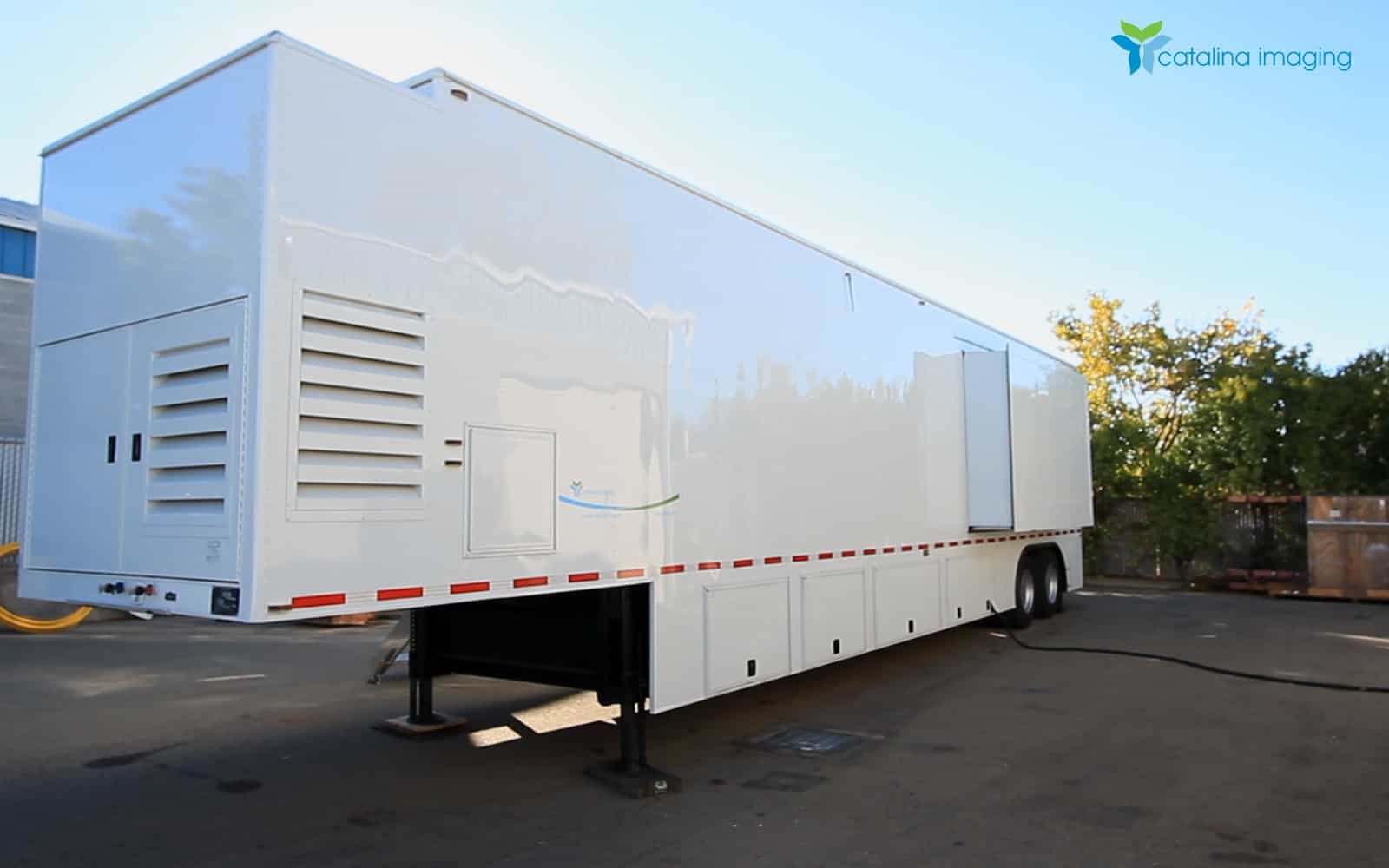
With the experts’ opinion and published studies regarding the clinical consequences of using a CT scan for TAVR procedures, the next big thing for clinics, small hospitals, and private imaging centers is the mobile CT scan.

A network of facilities can lease a single mobile CT scan which further reduces the upfront cost in owning one. Aside from lesser cost in initial investment, leasing or renting a mobile CT scan assures that the patient will have all their needs at the center’s disposal.

In some cases, the patient has to transfer to another facility to obtain a CT scan procedure and results. This is avoided when the facility has leased a mobile CT scan.

Using a mobile CT scan reduces the backlog when the machine has a dysfunction, it can be replaced with another leased unit in the soonest time possible. This avoids complications in patients with urgent care needs.

In conclusion, the convenience and efficiency of mobile CT scans can save a lot of time and money for a facility, in turn, better patient care and human resource improvement will be invested more of.

(Images of the aorta taken from CT scans using a GE Revolution HD. )

The GE Revolution Discovery HD: Now Available from Catalina Imaging

The GE Revolution Discovery HD: Now Available from Catalina Imaging

The latest addition to the Catalina Imaging fleet of mobile CT scanners is the GE Revolution Discovery HD.

With the GE Revolution Discovery HD, you get better full-body tomography exams with superb quality. The  Revolution HD features Smart Technologies, a suite of intelligent CT tools designed to help you achieve your imaging goals with improved efficiency and expanded applications, all while delivering diagnostic confidence with lower levels of radiation.

The cutting-edge CT technology featured on the GE Revolution Discovery HD includes:

  • Smart Dose with kV Assist provides automated kV and mA recommendations allowing users to tailor protocols to each patient, clinical indication, and each scan area.
  • Smart Cardiac with SnapShot Assist helps you optimize cardiac scanning for individual patients based on their specific parameters.
  • Smart Spectral with GSI Assist helps you optimize GSI protocols to your patient and clinical needs.

Why You’d Want The GE Revolution Discovery HD

You and your patients continue to demand a lower dose, but not at the expense of diagnostic image quality. Revolution HD delivers on both imperatives with a number of clinical benefits:

1.  Confidence boosting clarity

Spatial and temporal resolution, signal-to-noise ratio, low-contrast detectability, and artifact reduction are all fundamental to CT image quality.

The Revolution HD offers a true diagnostic breakthrough with a best-in-class spatial resolution of 0.23 mm across the full two-meter scan length, 29 msec of temporal resolutions, and Smart MAR is designed to reveal anatomic details obscured by metal artifacts, helping clinicians diagnose disease with greater confidence.

2. Low dose made possible by iterative reconstruction

Typically, lowering dose has increased noise and image artifacts, creating a difficult balance between higher image quality and lower dose. To help you overcome these challenges and keep you on the cutting edge,

The Revolution HD offers three advanced iterative reconstruction technologies, ASiR (standard), ASiR-V (option), and Veo (option).

3. Gemstone™ Spectral Imaging (GSI) helps you make a more confident diagnosis

GSI enables you to take CT beyond classical anatomical assessment to quantitative tissue characterization and advanced functional imaging.

More than 15 applications are now in routine use including oncology, cardiology, neurology, spine, urology, and musculoskeletal.

(To learn more about GSI clinical benefits, visit the GSI image gallery and the GSI education center.)

4. Best-in-class cardiac CT

Revolution HD has a best-in-class cardiac CT spatial resolution. At 18.2 lp/cm, the system provides up to 66% greater spatial resolution than comparable systems.

Offered as an option, SnapShot™ Freeze is designed to reduce blurring artifacts due to motion in coronary vessels that cannot be addressed by gantry speed alone.

Providing up to 6x improvement while maintaining high spatial resolution, the reduction in motion artifacts is equivalent to a 0.058s equivalent gantry rotation speed with an effective temporal resolution of 29 msec.

5. Focus less on the system and more on your patients

The Xtream display prominently shows the patient name, making exams more personal. It also includes a number of educational videos that explain CT procedures or can be used as a distraction technique for younger patients. In addition, with one-stop ED mode, you can select and confirm patient, protocol and scan settings at the gantry.

Canon Medical Launches CT Solution in Answer to COVID-19

Canon Medical Launches CT Solution in Answer to COVID-19 | Catalina Imaging

Last April 29, Canon Medical Systems USA, Inc, launched “deployable CT solutions and rapid decontamination” to help hospitals that are screening and caring for patients suspected to be positive or confirmed to be infected with COVID-19.

This may also be helpful in the isolation and imaging of patients who may be suffering from other viral infectious diseases.

Completely Capable System

This new deployable system involves the Aquilion Prime SP. This CT system offers the following benefits, among many others:
● Hit multiple anatomical areas within one breath-hold and one contrast injection
● Give healthcare workers access to the patient from the front and rear of the gantry
● Can be installed in most existing CT rooms

According to Canon Medical, the Aquilion Prime SP system “can generate 160 unique slices per rotation with 0.35-second scanning, reducing the time required to perform studies for compromised patients, and also increase throughput.” This makes the system useful even in emergency cases.

In Canon Medical’s new offering, the Aquilion Prime SP CT system can be used in a modular or mobile footprint with a rapid decontamination tool. This assures rapid imaging, safe handling of patients, complete isolation (even from other hospital staff), and necessary decontamination.

Cleaning and Decontamination in Minutes

According to Diagnostic Imaging in an article written by Whitney J. Palmer, “In most cases, based on industry guidance, a CT scanner has been decommissioned for roughly an hour between scanning patients to minimize the likelihood of viral transmission.”

An hour can already be the length of a church service. If we recall the story of Patient 31, after she attended two services at her church in South Korea, she ended up infecting 1,160 people as of March 20.

Hospitals and healthcare workers do not have the luxury of an hour to make sure their CT scanner is ready for another patient — not in a time like now with its demands.

Rapid UV-C Decontamination

Canon Medical addresses this with its system that can be decontaminated in minutes, with the use of an automated ultraviolet light system. “Imaging infectious disease patients is not a new phenomenon for medical providers,” said Erin Angel, managing director, CT Business Unit, Canon Medical Systems USA, Inc, in a statement.

“But our customers are facing an unprecedented number of potentially contagious patients,” he continued. “Our deployable CT offerings with the addition of rapid UV-C decontamination will offer providers a unique solution to help improve their workflow and safety as they image infectious disease patients.”

Least Chances of Contamination

Simply put, Canon Medical’s decontamination system is an automated UV-C technology that significantly reduces bacteria, spores, and viruses. It is effective against a variety of advanced viral infectious diseases. It is readily available for new CT solutions as well as existing imaging suites.

The system’s multiple automated UV-C emitters work together to provide decontamination of the room in minutes, a big factor especially with a virus as quick to spread as SARS-CoV-2 (this is the official name that the World Health Organization gave the coronavirus or the virus that causes COVID-19).

Experts Warn Radiology Practices To Prepare For a Coming Surge

Experts Warn Radiology Practices To Prepare For a Coming Surge | Catalina Imaging

Radiology experts are expecting a surge in demand in the coming months once routine cancer screenings finally resume.

The COVID-19 crisis has forced radiology providers to postpone all nonurgent cancer imaging across the country. But once things have settled down, however, practices are likely to be overwhelmed by the sudden influx of procedures. 

As early as now, experts recommend that hospitals and clinics should prepare for the coming surge. An editorial published by the Radiological Society of North America(RSNA) and the University of Michigan has explored such scenarios and what’s needed in terms of adequate preparation.

The editorial, Radiology: Imaging Cancer, was recently published in Michigan Medicine. Gary Luker, MD is the editor of the journal, while Adeline Boettcher, Ph.D. of the RSNA is its scientific editor.

“Even with proposed modifications to availability of scanners and examination time, most institutions already operate imaging equipment at near capacity with appointment slots during evenings and weekends,” warned Luker and Boettcher. “Trying to reschedule several months of imaging appointments likely will result in prolonged (weeks to months) wait times, which may deter many patients.” 

Other key points of the editorial:

  • Institutions face dual challenges of maintaining operations during stay-at-home safety orders and planning for a future that will not resemble our past “normal”.
  • Fear of hospitals and loss of insurance from unemployment likely will be obstacles to cancer screening and other non-emergent imaging studies.
  • A backlog of postponed and rescheduled imaging studies will create a surge that taxes capacities of scanner time and personnel.
  • The economic impact of the pandemic threatens current and future support for imaging scientists and research activities in cancer imaging and image-guided therapy.

Radiology - Woman undergoes CT Scan

Physicians generally estimate that regular appointments might start to resume during the month of May. Which wouldn’t be a problem, except that there will be a continued need for social distancing and additional safety protocols. 

To complicate matters even further, layoffs of staffers and patients losing their health insurance add to prolonging the amount of time to get back to normalcy– probably taking as long as 6 to 12 months, noted Luker and Boettcher.

According to experts tapped by Luker and Boettcher for the editorial, patients are most likely to continue to receive routine cancer screenings, once the pandemic ends. These same experts, however, pointed out that some may remain fearful of going out into public, especially individuals undergoing chemotherapy who are at increased risk of severe COVID-19 infection. Additionally, others who are in remission may choose to avoid follow-up imaging.

“In this regard, it will be important that clinics emphasize that they will take every precaution necessary to keep these patients safe and protected during their appointments,” reminded the authors, suggesting additional measures such as dedicated entrances and traffic patterns for specifically for immunocompromised patients.

Meanwhile, in countries where hospitals have already resumed normal cancer screenings (such as in China), the vast majority of patients and procedures are likely still in a holding pattern. Luker and Boettcher have suggested that telehealth appointments are one way to get subjects to “reengage in cancer imaging studies.” Reminders via email, mobile messaging, and social media applications might also prove helpful. 

Whether it’s routine procedures or medical studies, practices should expect social-distancing policies remain in force into 2021. Even when operations start returning to some semblance of normalcy, hospitals and clinics should prepare for this reality to not just protect staff and customers, but to also provide for additional sanitation measures such as more frequent cleaning for imaging rooms and equipment. 

(Source: Radiology Business)

Are CT Scanners Useful in Diagnosing COVID-19?

Are CT Scanners Useful in Diagnosing COVID-19? | Catalina Imaging

Recent literature in radiology suggests that CT scanners may play an important role in the diagnosis of COVID-19.

In a recent article published in The Lancet, a team of researchers has observed that almost all of the confirmed COVID-19 cases under their care also had CT findings of pneumonia.

These medical researchers, working with coronavirus patients in Wuhan, China, have found that 97% of their 1,014 patients with RT-PCR-confirmed diagnoses also had observed lung opacities on CT— a notable manifestation of pneumonia. In their report, the medical researchers have concluded that “CT imaging has high sensitivity for diagnosis of COVID-19”.

This is particularly welcome news, especially with shortages of real-time RT-PCR (reverse transcription-polymerase chain reaction), so far one of the most effective and accurate laboratory methods for detecting, tracking, and studying the coronavirus.

Further Investigation Needed

Meanwhile, another group of researchers has had less optimistic observations.

A team that had reviewed the CT scans of 112 cases of RT-PCR-confirmed COVID-19 from the Diamond Princess cruise ship reported that only 61%– or less than two-thirds– of cases exhibited pneumonia. Furthermore, 20% of symptomatic patients had negative CTs.

So while CT scans do show promise in the diagnosis of COVID-19, these results should not be overstated, cautioned The Lancet.

“The CT findings studied (e.g., ground-glass opacity, consolidation, etc.) are not specific for COVID-19. Similar results would probably be found if CT were used during an influenza epidemic, for example.”

Bringing A.I. Into The Picture

Meanwhile, a medical devices company based in Huizhou, China is exploring the possibility of using an AI-powered imaging solution to aid in the detection of COVID-19

Huiying Medical, a member of Intel’s AI Builders program, claims to have developed the solution’s underpinning algorithms based on data from CT chest scans from over 4,000 coronavirus cases.

Huiying’s solution analyzes what’s known as ground-glass opacity (GGO) in the lungs, which indicates a partial filling of air spaces, as well as other indicators that inform a probability of suspected COVID-19 infection.

Huiying asserts that its solution should be useful in regions of the world without access to real-time RT-PCR. It only takes a day to install and only 2-3 seconds to process CT studies with 500 images.

Moreover, it has a claimed 96% novel coronavirus pneumonia (NCP) classification rate, and it’s designed to work either in the cloud or on-premises.

Through a partnership with Huawei, Huiying has already reached out to many other health professionals and institutions. The new AI-enabled imaging solution is now being used in over 20 hospitals, including those in Ecuador and in the Philippines.

Promising, But Needs More Work

The Lancet stresses that, despite its limitations, RT-PCR is still the accepted standard and only positive in patients who are infected with severe acute respiratory syndrome coronavirus 2.

While advancements in CT technology and research shows promise, the medical journal cautions using published guidelines regarding the use of CT imaging– at least for now.

CT findings in patients with COVID-19 are seen with numerous pathogens and in many non-infectious aetiologies. “Using CT diagnostically is not known to provide clinical benefit and could lead to false security if results are negative,” warns Michael Hope of The Lancet.

“If COVID-19 is suspected, patients should be isolated pending confirmation with (multiple) RT-PCR tests,” he stressed, “or until quarantine has lapsed. The results of a CT scan do not change this.”

Recommendations From The CDC and the ACR

Currently, the Center for Disease Control (CDC) does not currently recommend CXR (chest radiographs) or CT to diagnose COVID-19.

The American College of Radiology (ACR) has its own supporting recommendations for the use of computed tomography (CT) and chest radiography for suspected COVID-19 infections.

Even if radiologic findings are suggestive of COVID-19 on CT, confirmation with the viral test is still required. As such, viral testing remains the only specific method of diagnosis.

In the meantime, until more widespread COVID-19 testing is available, some medical practices are requesting chest CT to inform decisions on whether to test a patient for COVID-19, admit a patient or provide other treatment as an interim measure.

As such, facilities may consider deploying portable radiography units in ambulatory care facilities for use when CXRs are considered medically necessary. The surfaces of these machines can be easily cleaned, avoiding the need to bring patients into radiography rooms.

While locally constrained resources may be a factor in decision making, a normal CT should not dissuade a patient from being quarantined or provided other clinically indicated treatment when otherwise medically appropriate.

Mobile CT Scanners By Catalina Imaging

Catalina Imaging is committed to providing the highest quality solutions by specializing in mobile CT scanners using state-of-the-art Siemens, GE, and Toshiba/Canon technology.

The CT scanners we use in our trailers are the best available from GE, Toshiba, and Siemens. Depending on your needs you’ll receive GE’s Lightspeed VCT64 with ASiR, Toshiba’s Aquilion 16 MultiSlice Computerized Tomography Scanner, or one of our Siemens mobile CT units.

For details and more information about our mobile CT scanners, give our Catalina Imaging team a call today at (844) 949-1664.

Sources

  1. The Lancet
  2. The International Atomic Energy Agency
  3. Venture Beat
  4. American College of Radiologists


Radiologist Burnout

radiologist examining x ray

Burnout is almost twice as common among physicians compared to other US workers. The burnout rate varies widely by specialty and radiologists are right at the average with 45% reporting symptoms of burnout.


percentage of radiologists feeling burned out


According to the Medscape Radiologist Lifestyle, Happiness, & Burnout Report 2019, only 25% of radiologists consider themselves to be very or extremely happy in their jobs, while 53% are very or extremely happy outside their jobs. This discrepancy between workplace and personal happiness is startling, and it may reflect the burnout experienced by many radiologists.

What is Burnout?

According to the Mayo Clinic, burnout is a type of work-related stress that is “a state of physical or emotional exhaustion that also involves a sense of reduced accomplishment and loss of personal identity.”
This type of job-related burnout may have devastating effects on both your physical and mental health. Radiologists experiencing burnout may feel a lack of motivation for their jobs, which may lead to a lack of compassion or care when working with patients. Eventually, they may even struggle with remembering why they entered the medical field.

Signs of Burnout

  • There are some specific signs to look for to determine if you experiencing burnout. Ask yourself the following questions to see if this may be happening to you.
  • Have you become cynical about your work or impatient or critical with co-workers or patients?
  • Do you have difficulty concentrating and being productive?
  • Have you experienced physical symptoms, such as a loss of energy, a change in sleep habits, unexplained headaches, intestinal problems, or other physical problems?
  • Do you feel disillusioned or dissatisfied with your work?
  • Do you use food, alcohol, or drugs as an escape mechanism?

Causes of Burnout for Radiologists

It may be common for radiologists to experience a feeling of disconnection in the workplace. Due to the nature of the job, they may not have many opportunities for interaction with patients or their referring physicians.

According to Cheri L. Canon, MD, FACR Chair of Radiology at the University of Alabama School of Medicine, the reading room can also be an isolating environment, which may lead to radiologists feeling cut-off from their patients and colleagues.


A variety of factors contribute to burnout, including the following:

  • Working excessive hours or lack of work/life balance.
  • Lack of respect from administrators and co-workers.
  • Feeling like a cog in a wheel.
  • Lack of control.
  • Bureaucratic tasks, such as excessive paperwork.
  • Insufficient compensation.
  • Emphasis on profits over patients.
  • Government regulations.
  • Increase in digital recordkeeping, such as EHRs, which may be timeconsuming and distracting.
  • Lack of respect from patients.


what contributes to radiologist burnout


How do Radiologists Cope with Burnout?

According to the Medscape Report, 54 percent of radiologists use exercise to cope with burnout, 41% talk with family members or close friends, 36% sleep, and 35% listen to music. These are all healthy techniques that may be helpful in dealing with burnout.

Unfortunately, some radiologists engage in more destructive behaviors to cope, such as isolating themselves from others, eating junk food, binge eating, drinking alcohol, smoking, and using marijuana or prescription drugs. These coping mechanisms may lead to health problems and make the situation worse.


How to Prevent and Overcome Burnout

Individual physicians have varying levels of how much stress they can tolerate, and what one sees as burnout, another may see as a normal amount of stress or fatigue. It’s important to remember that not everyone handles the physical, emotional, or mental stress of burnout in the same way, but there are some things you can do to help alleviate the stress of burnout.

  • Find and address any sources of job dissatisfaction.
  • Work to develop self-care habits that will reduce stress and improve your physical, mental, and emotional health, including eating healthy foods, staying active, and getting adequate sleep.
  • Make sure you are properly balancing your work with the rest of your life by taking adequate time off for vacation or personal time and spending time with your family and friends.
  • Remember why you chose your field and what accomplishments you have achieved.
  • Find support when needed, either through colleagues, a support group, or close friends and family members.

The Role of Medical Facilities and Organizations

Medical facilities and organizations may also help prevent burnout by creating a culture that provides radiologists and other medical professionals with the support they need to be effective in their jobs and satisfied with their work. Below are some things organizational leaders may do to help reduce burnout.

  • Encourage an atmosphere of community and connectivity to reduce the isolation many radiologists and physicians may be experiencing.
  • Model behaviors that encourage wellness among staff members.
  • Advocate for ways to reduce stress and encourage overall wellness.
  • Help eliminate the stigma of burnout by encouraging staff members to find help if they need it.
  • Pay attention to individual staff members to see signs of potential burnout and prevent it.
  • Measure data and use it to monitor progress.
  • Consider creating a wellness center or forming a committee to address burnout prevention and job satisfaction.
  • Prioritize patient care and physician satisfaction.
  • Coordinate goals, roles, and processes so they are aligned.
  • Create an environment where staff members are all respected and valued.
  • Acknowledge accomplishments to boost morale.
  • Improve efficiency by consolidating tasks and eliminating unnecessary work.
  • Consider implementing innovative AI technology to streamline workflow, automate routine tasks, and improve reporting, so radiologists can focus on consistently providing accurate reports and recommendations as efficiently as possible.
  • Implement PACS and EHR patient context integration to improve efficiency and save time when accessing clinical data.


preventing and reducing radiologist burnout


If you think you may be experiencing burnout, pay attention to how you feel, and address your concerns so you can get the help you need. It’s important to take care of yourself so you can be effective in your job as you take care of others. Remember the reasons why you chose this profession, and remind yourself of your achievements and accomplishments as you take the necessary steps to reduce your stress, improve your wellness, and become passionate about your work again.

Sources: 

https://jamanetwork.com/journals/jamainternalmedicine/fullarticle/1351351

https://www.mayoclinic.org/healthy-lifestyle/adult-health/in-depth/burnout/art-20046642

https://www.medscape.com/slideshow/2019-lifestyle-radiologist-6011151#1 

https://www.ncbi.nlm.nih.gov/pubmed/26768546 

https://www.thedoctors.com/about-the-doctors-company/newsroom/the-future-of-healthcare-survey

https://www.radiologybusiness.com/topics/leadership/radiologist-burnout-are-we-done-yet 

https://acrbulletin.org/topics/practice-management/1447-strategies-for-burn-out

https://www.nuance.com/healthcare/campaign/ppc/improve-radiology-efficiency-burnout.html?cid=7010W000002SiTjQAK&cn=DHD-AR-FY2019-Efficiency&Burnout-Diag-PPC&ls=CPC&rs=google&pd=Y&gclid=CjwKCAiA6bvwBRBbEiwAUER6JTA0sMMAc7w1Z2uJAP86a-feuqfdHV5BGlriFzj8CqhwQ4VYaVHEKBoCqFAQAvD_BwE

https://www.radiologybusiness.com/topics/imaging-informatics/pacs-emr-integration-saves-radiologists-time-improves-emr-utilization

Mobile CT Scanner Rental and Lease Guide

Mobile Imaging Electrical Trailer Schematics

In this article, we combine our 50 years of combined experience as mobile imaging engineers with the feedback of thirteen radiology managers to provide you with the most thorough article available on renting a mobile CT scanner.

border top
See what the inside of a trailer looks like with this video:

border bottom


Why A Mobile CT Scanner?

Renting, leasing, or purchasing a mobile CT scanner can be an excellent interim solution for a variety of circumstances.

The most common call we receive is from the radiology department of a hospital or clinic whose CT machine has gone down. The following are additional situations where an interim solution is beneficial:

  • Building/department renovations
  • Testing a market
  • Low traffic at multiple locations
  • Research studies
  • Rural visitations
  • Avoiding large capital expenditures

What Does It Cost To Rent A  Mobile CT Scanner?

Please email info@catalinaimaging or give us a call for a Mobile CT Quote

Prices will vary depending on the quality of the unit, the make and model of the CT scanner, and whether you’ll be taken care of by an OEM service provider.

Quality Evaluation

The quality of the unit includes:

  • Age of the trailer encompassing the mobile imaging equipment
  • How well kept up the unit is (fixes, wear and tear, etc)
  • Aesthetics (Is it cold and bland or have the look and feel been considered?)
  • Comfortability (Is it a tight fit or does the layout allow the patient and nurse to move around comfortably?)

The most common equipment manufacturers are Toshiba, GE, and Siemens. All offer different makes and models of mobile CT units.

Additionally, the unit in the trailer could have been acquired new or refurbished.

Toshiba Mobile CT Scanner
Above: Toshiba Aquilion 64 MultiSlice CT Scanner in a Mobile Trailer

A major deciding factor for the radiology managers we interviewed was the brand of the equipment. A hospital or clinic needs the interim CT scanner to tie into its existing technology.

Aligning technologies also makes training staff much easier since they will already be accustomed to the user interface and user experience the brand provides.

The third major component is who will be providing your ongoing service.

OEM Provider vs Third-Party Provider

As one of our study’s more experienced participants put it, “It’s expected that problems will arise when leasing a unit.” This does not mean issues are common. He acknowledges the intricacy of each project.

While third-party service providers can cost as much as 30% lower, more experienced radiology managers know that this can come at a cost.

No matter how you slice it, going with a third-party service provider raises the risk of receiving lower-quality service.

Downtime is very costly and can be a huge headache. The number one and number two priorities of our radiology managers when evaluating what mobile CT service provider to use were directly related to resolving downtime issues. These included customer service and service availability.

If the engineer sent to your site is undertrained or inexperienced this could end up costing thousands more than it needed to, not to mention the stress that comes with dealing with upset patients or higher-ups.

Another consideration; some companies offer 24/7 service with their rental and leasings. Be sure to find out if the companies you’re looking at do.

Extra Costs to Consider

Each mobile imaging company will have its way of handling these extra costs, so it’s important to set clear expectations of what you are responsible for.

Some costs to consider include:

  • A security deposit
  • Site help, pre-planning, and set-up
  • Transportation and delivery
  • Applications training
  • De-installation
  • Cleaning fees

Planning For Your Unit

Because a mobile CT unit is such a specialized piece of equipment the logistics involved in planning and preparing the site are extensive. If you’re working with a company that knows what they’re doing and you make yourself available to work with them then the process can be smooth. The further in advance you plan though, the better.

“Mobile
Above: A trailer that holds the mobile CT scanner.


Some logistics to consider include the ability of the truck to maneuver the trailer in place, citing issues such as the levelness and condition of the ground, weather conditions, and electrical requirements.

The pad the unit will be set on needs to be able to support 30 or more tons of weight. It also needs enough space for both the mobile CT trailer and the transportation of patients in and out of the trailer. Both the size of the area needed and the dimensions of the trailer will vary depending on the company.

Mobile CT Trailer Guide

Above: An overhead view of a mobile CT trailer and the area required for it.

Ongoing Maintenance And Customer Service

As we mentioned earlier, this is a big deal for the radiology managers we spoke with. You’ll want to make sure there are multiple points of contact for situations involving both the trailer holding the CT equipment and the CT equipment itself. If your company is an OEM service provider then your point of contact for the mobile imaging equipment will likely be the OEM.

De-Installation

Once you’ve scheduled a time for the service engineer to come out and unplug everything you’ll want to make sure you remove all patient data. You’ll also need to remove all items you brought into the trailer and you may be responsible for final cleaning.

Conclusion

When searching for a mobile imaging service provider the top three priorities of the radiology managers were customer service, availability, and price, in that order.

When looking for your own company try to run through the checklist of questions provided in this article to make sure they’re up to providing the highest quality service. Renting a mobile CT scanner can be a smooth experience if done with the right company. We hope this article brought some clarity about what that company should look like.


Looking for a mobile CT? We love helping new customers!

Contact us today: (844) 949-1664

Our Latest Addition To The Fleet: GE 750HD High Definition Imaging CT

GE 750HD CT Scanner

In our pursuit to always carry the most state-of-the-art technology for our clients, we have added a new and improved GE unit to our fleet.

High Definition Imaging-GE’s high-performance system combines the benefits of the Gemstone Detector, liquid bearing tube, spectral imaging and 128 slice capability to produce the industries best spatial and contrast resolution at 230 micron spatial resolution.

Gemstone Spectral Imaging-Innovative technology which uses fast kV switching dual energy acquisition coupled with fast sampling, low afterglow and high light output scintillator detector to produce near perfectly registered dual energy.

Performix HD Tube-GE’s patented MX 240 Perseus high-performance liquid bearing tube technology produces superb image quality when combined with the Gemstone detector, while enabling Smart Technologies to deliver efficient dose modulation and longer tube life.  

GSI Cardiac-Provides enhanced coronary assessment with ability to alternate two kVp energies at .25msec for unprecedented temporal registration.  

Additional features;

  • Dynamic Z-axis tracking-low dose in helical scanning
  • GOC-6.66 Console
  • Cardiac and Neuro acquisition
  • Xtream Recon
  • Organ Dose Modulation-Automatically protect sensitive organs (eyes etc.)
  • GSI SnapShot Pulse-Intelligent Motion Correction for high heart rate cardiac CT
  • Smart MAR-Advanced metal artifact reduction technique

Call us today at (844) 949-1664 to learn more about how the GE 750HD can work for your hospital’s needs.

MRI or CT Scan: What’s the Difference?

Female undergoing CT Scan

CT scans (also known as CAT scan) and MRIs are two of the most commonly performed imaging techniques to help doctors diagnose, and sometimes treat, injury or disease. A CT scan (computed tomography) employs x-rays while MRI (magnetic resonance imaging) uses a magnetic field and radiofrequency pulses; both techniques are able to provide a look at the internal structure of the body.


Given that each test seemingly provides similar results, you might wonder why your doctor would choose one instead of the other.


MRI

Doctor standing in front of an MRI machine


As the name suggests, MRIs use magnets to generate images. These aren’t your everyday refrigerator magnets though. MRIs use huge superconductive magnets which have a greater magnet field strength thousands of times stronger than that of the earth’s. MRIs do not use ionizing radiation and are able to image the brain, spine, organs, bones/joints, and soft tissues quite nicely. So MRI must be better than CT right? The answer is not so simple.


The machinery needed for MRI is more expensive and the superconductive magnet requires significant safety precautions so they are not as readily available as CT. Additionally, the process of producing an MR image is inherently slower and the patient must be able to remain completely still. MRIs can take up to 30 minutes, sometimes longer, depending on the body part and the number of pictures needed to be acquired. Therefore MRI is often not the best choice in the setting of emergencies, bleeding, trauma, and in patients who are claustrophobic or are unable to remain still for long periods of time.   


Because MRIs use a powerful magnet, patients who have internal metal, medical devices, or certain piercings may not be eligible for an MRI. Internal devices such as defibrillators, pacemakers, stimulators, or shunts may be affected by the magnetic field and cause the device to malfunction (of note medical device companies are currently designing devices which are MRI safe, several of which are already on the market). Piercings and metallic foreign bodies can heat up in the magnet resulting in burns. Most radiology departments complete an extensive screening questionnaire to identify any potential hazards. 


If you have a medical device be sure to save the manufacturer’s card and information so it can be fully reviewed by radiology staff prior to your MRI. If your device is not safe for the MRI, the radiologist may recommend a different type of imaging. Additionally, be sure to remove piercings and alert the radiology staff to any metal that may be on or inside your body.


CT Scans

Doctor standing next to CT scanner


CT scans are readily available at most hospitals and can be obtained rapidly, so are commonly used in emergency situations. In cases where a CT scanner hasn’t been made available, a mobile CT scanner can be requested for these emergencies. The patient lies on the table which quickly moves through the CT tube and images are obtained.


CT scans can produce images of the bones, chest, internal organs, digestive tract, and blood quite well so is commonly used to diagnose head injuries, abnormalities in the chest or abdomen, fractures, or internal bleeding.  In the nonemergent setting, CT scans can be used for imaging heart and lung disease, abdominal abnormalities, bones/joints, and cancer.


CT scan of healthy lungs


Additionally, because CT scans provide an assessment of patients anatomy, surgeons may opt to obtain a CT scan for surgical planning (like a road map).


CT Scans Expose Patients to Radiation

Human graph of average radiation exposure


Now let’s address the elephant in the room, radiation. CT scans use x-rays that pass through your body to generate images. X-rays are a type of ionizing radiation, which means they have enough energy to create ions of the material they pass through. When cells inside the body (i.e. the patient) are exposed to ionizing radiation, the DNA inside can become damaged and the cell can then become cancerous. 


Don’t worry too much. You are actually exposed to low levels of radiation that exist naturally in the environment every day and generally speaking, your cells do a pretty good job of DNA repair. For comparison sake, an average person is exposed to 3 mSv of radiation annually just from the environment, while an average CT scan is between 2-8 mSv. The good (and bad) news is that there is no radiation dose or threshold point at which one will develop cancer, but most scientists agree that the risk of cancer increases as radiation dose increases.


When you and your doctor decide for you to get a CT scan, you are deciding that the risks of the radiation are less than the disease or injury you are looking to diagnose. 


Conclusion

CTs and MRIs each have their own unique risks and benefits which need to be applied to an individual patient to answer a clinical question. When used appropriately both have proven to be a safe and effective means of diagnostic imaging. If your doctor recommends you receive a CT or MRI, feel free to ask why they ordered one versus the other. Also, be sure to direct any additional questions to the radiology personnel prior to your exam.

Disclaimer: Please note the information provided in this article is a broad generalization and is designed for educational purposes only. This article does not constitute medical advice. It is recommended you consult with your physician with concerns or questions related to your specific medical condition and for additional recommendations.  

History of the Mobile CT Scan

patient using mobile CT scanner

The incredible imaging capacity of computed tomography (CT) has pushed the medical industry to staggering heights. Unfortunately, CT scans have yet to help out the majority of medics in the battlefields, ambulance crews rushing to emergency situations, and even emergency room doctors. This is because conventional CT scanners are practically behemoths, weighing roughly 4,000 kilograms as well as requiring high-voltage capacities to power massive cooling systems and climate-controlled radiology rooms.

mobile CT scanner 16 slide system

Above: An image produced from a 16 slide CT scan system.

The lack of convenient and quick access to CT scans, especially when dealing with stroke or injuries involving brain trauma, has become a critical issue in the medical industry. Despite the continuous advancements in this realm of imaging technique, the immobility of CT scans has been a pain point that needs to be addressed as soon as possible. This is where the portable CT scanner comes in.

Around the 1990s, two types of CT scanners emerged. One is the “fixed” CT scanner, which refers to the huge machines still found in the majority of hospitals across the globe. The second is the mobile CT scanner, or the “portable” CT machine, which is lightweight and can be easily transported.


History of the Mobile CT Scanner

While there is no specific time and date that could pinpoint the first time a portable CT scanner was used, in 1949 came a surge in demand for “mobile health care.” Around the 1970s, the world’s first mobile CT scanner was launched by Medical Coaches Inc. via their founder Ian Smith’s deal with Peru.

This mobile CT scanner focused primarily on head scans and attempted to provide cross-sectional images of the heart as well. Apart from this, the mobile healthcare unit at the time also provided ultrasounds for pregnant women to evaluate the status of their hearts, gallbladders, breasts, and livers.


What are mobile CT scanners used for nowadays?

Throughout the years, the demand for a mobile CT scanner has increased exponentially due to the influx of incidents where medics have failed to swiftly provide care to stroke patients. These incidents paved the way to the study of utilizing and funding mobile stroke units (MSUs) in 2003. In 2008, the first-ever clinical application of an MSU was conducted at Saarland University in Germany.

mobile CT scanner used for MSU mobile stroke unit

Above: A modern MSU.

The initiative was rooted in the idea of “bringing the hospital to the patient,” which eventually decreased the waiting period from the initial distress call to therapy. More importantly, the presence of a portable CT scanner in these MSUs ensured that the professionals could treat patients quickly and accurately during emergencies.

Gradually, the need for MSUs featuring computed tomography equipment inspired 20 more sites across the globe. In January 2014, Houston’s Frazer Ltd designed and released the first MSU in the United States. Basing its setup on the recommendations of neurologists, the company incorporated a portable CT scanner in its hospital on wheels to drastically cut down on treatment time. By 2016, New York’s Presbyterian Hospital became the first to field an MSU on the East Coast and reached a total of three functioning MSUs by 2018.

Another hospital that’s taking advantage of MSUs and portable CT scanners is the University of Tennessee Health Science Center. Aside from the mobile CT scanner, the 14-ton ambulance is also equipped with tools that can infuse early fluids to their stroke patients as well as dye blood vessels to figure out the kind of stroke. Given that Tennessee has been identified as a “stroke belt,” this technology has been a life-changer for a lot of people.

The Memphis MSU staff takes only 13 to 14 minutes to provide treatment to their patients, which covers their response to the scene up to the application of medications to the veins – a far cry from the 40 to 50 minutes these tasks take in emergency rooms.

Neurologists have been hoping for more portable CT scanners, particularly in intensive care units. The availability of a mobile CT scanner on hand would exponentially increase their efficiency, eliminating the need to transfer patients and all medical personnel to the location of the humongous fixed CT scanner.


Final thoughts

Computed tomography has evolved in an impressive way over the past 40 years. While it might be nearly impossible for the world to reach the advanced level of technology presented in Star Trek, where doctors can simply wave their wands to instantly diagnose their patients, this rapidly developing niche in radiology seems to be getting there.