Mobile CT Scanners vs. Portable CT Units: What’s the Difference?

Mobile CT Scanners

CT scanners are important medical devices that are used to produce detailed images of the inside of the body. While both mobile CT scanners and portable CT units can be used for this purpose, there are some key differences between the two types of scanners.

In this article, we will compare the size, mobility, and versatility of mobile CT scanners and portable CT units, and discuss the pros and cons of each. We will also explore the various uses of these scanners, including in emergency situations and in remote or rural areas where access to traditional CT scanners may be limited.

At Catalina Imaging, we’re committed to providing the highest quality solutions by specializing in mobile CT scanning technology. Contact us today at info@catalinaimaging.com or call us at (844) 949-1664!

What makes Mobile CT Scanners vs. Portable CT Units so different?

They use different systems

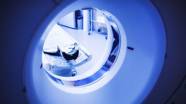
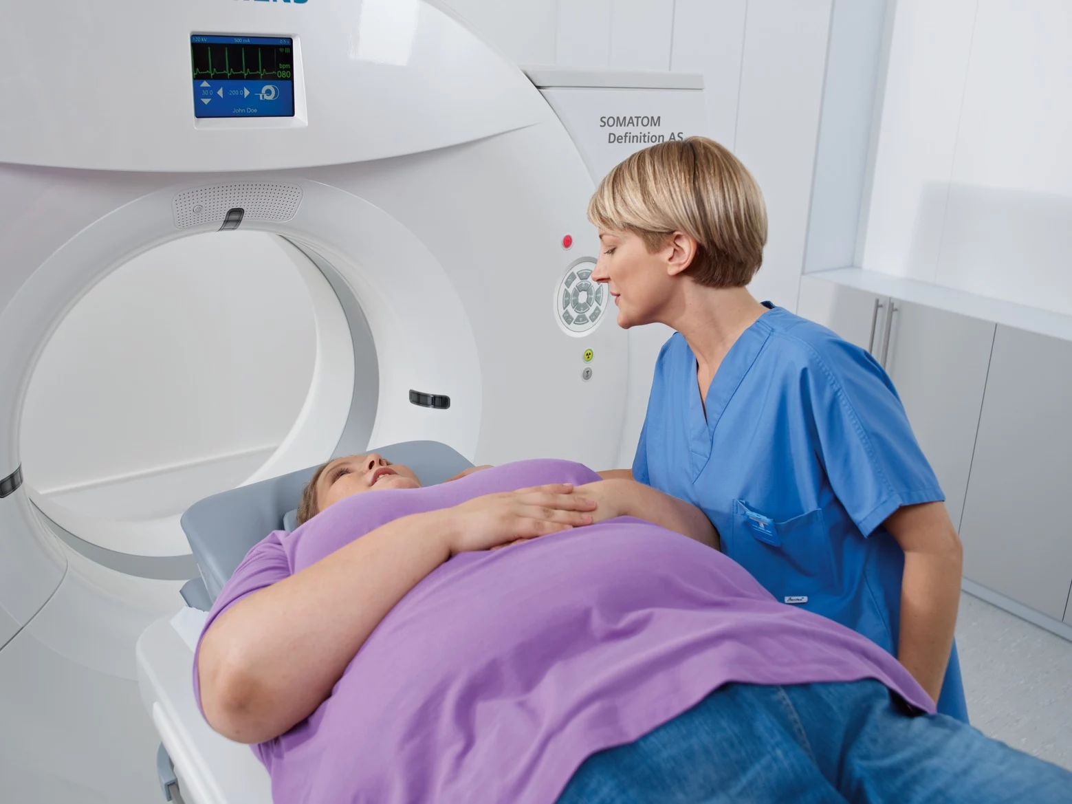
Mobile CT scanners have a rotating gantry that allows them to go around the patient during the scan. The gantry is a large, stationary cylindrical tube that supports the x-ray source and detector array. These components work together to produce detailed images of the inside of the body.

The x-ray source emits a series of x-ray beams that pass through the patient’s body and are detected by the detector array on the other side. The resulting data is used to create detailed cross-sectional images of the body. They offer high resolution and detail, making them particularly useful for visualizing organs such as the heart, kidneys, and liver.

Portable CT units, on the other hand, use a single x-ray source and detector to produce detailed images of the inside of the body. The x-ray source in a portable CT unit is a small, focused beam of x-rays that is directed at the area of the body being imaged. The detector is located on the opposite side of the body and measures the intensity of the x-rays that pass through the body.

That being said, portable CT scanners are generally easier to use and require less training; however, they may not offer the same level of resolution or detail as mobile CT scanners.

They have different capabilities

Mobile CT scanners are typically mounted on a truck or trailer and brought to hospitals or other medical facilities for on-site imaging. They are generally larger, more expensive, and more complex than portable CT units and require a trained operator to perform the scan.

Additionally, mobile CT scanners are used for more specialized or complex imaging procedures. Portable CT scanners, on the other hand, are more commonly used in general diagnostic imaging and are used in remote locations, such as rural areas or developing countries. This is because they are small and easy to transport, allowing them to be moved easily from place to place where access to traditional CT scanners may be limited. For this same reason, they are also particularly useful in emergency situations.

Conclusion
Both mobile and portable CT scanners offer important benefits in terms of diagnostic accuracy and patient care and will continue to play a vital role in the medical field for years to come. Understanding the differences between the two devices can help healthcare providers choose the most appropriate device for a particular situation.

Let us help you find the perfect solution for your mobile medical imaging needs. Call us today at (844) 949-1664 or email us at info@catalinaimaging.com to learn how we can help your practice.

5 Common CT Scanner Issues and What To Do About Them

Common CT Scanner Issues

Computed tomography (CT) scanners are a vital piece of equipment in any medical imaging department. And after performing thousands of scans, it’s normal for scanners to experience some sort of issue. In the event that an issue does come up, typically an imaging engineer will be called in to diagnose and fix the problem as quickly as possible.


Common Ways That Your CT Scanner Can Fail and How to Avoid Them

To keep you better informed of the ins and outs of CT scanners, we’re going to talk about the most common problems you can expect when operating them, as well as what you can do to avoid them from ever happening. This way, you won’t have to worry about experiencing unnecessary downtime or ending up with an inferior scan.


At Catalina Imaging, we’re committed to providing the highest quality solutions by specializing in mobile CT scanning technology. Contact us today at info@catalinaimaging.com or call us at (844) 949-1664!


  • X-ray tube failure

When the X-ray tube fails or malfunctions, it can result in poor image quality or an inability to produce images at all. This usually occurs when there is an issue in the vacuum seal within the tube or damage to the tube itself. Other factors may also contribute to this issue, including age, wear and tear, and improper maintenance.


If an X-ray tube failure occurs, turn off the X-ray beam immediately to ensure the safety of the patient and the healthcare providers. The CT scan may need to be rescheduled or performed using a different type of medical imaging technology, depending on the severity of the failure and the unavailability of alternative equipment. The healthcare provider should work with the patient to determine the best course of action.


  • Calibration issues

Calibration is very important to ensure that the machine is working properly and giving accurate results. To calibrate a CT scanner is to scan an object called a phantom, with known radiodensity, to check whether the measurements given show the correct number of Hounsfield Units (HUs). 


This can be done manually or automatically, depending on what kind of calibration tool was included with your CT scanner purchase. If there isn’t any calibration tool included with your purchase, you will need to buy it separately.


  • Cloudy sensors and filters

Ambient temperature and humidity can affect the performance of your machine. This is especially important in humid environments, where water vapor can condense on the sensors and lenses of your device.


To avoid this problem, try to keep your machine at around 72 degrees Fahrenheit (22 degrees Celsius). And if you live in an especially dry climate, be sure to keep it out of direct sunlight.


Dust particles and other contaminants inside the air filter system can also be the culprit as to why your machine is malfunctioning. To prevent this, change out the CT scanner’s filters regularly (at least once every three months) and clean them regularly with compressed air, as well as soap-based cleaners (never use alcohol.) 


Additionally, it’s recommended to use HEPA filters for any rooms adjacent to where you plan on doing scans. These will capture most dust particles before they go anywhere near the machine’s sensitive components.


  • Too much image noise 

Multiple detector scanners are those that use two or more detectors to take x-ray images from different angles around the body. Each detector records only part of an image, which are all combined later on during processing. While these can produce better-quality results than single-detector scanners, they also tend to produce images with more noise. 


When you experience this, try to increase the radiation dose to improve the contrast resolution of a CT scan. This will reduce the amount of noise in the image, resulting in a clearer and more detailed image.


However, it is also important to consider the balance between the radiation dose and the required contrast resolution when determining the settings for the examination. Increasing the radiation dose may come at a cost, and it is necessary to weigh this against the need for a high-quality image.


  • Power surges

If a CT scanner were to experience a power surge, it damage your equipment. This is why it’s important to use surge protectors that are UL-tested and approved.


Furthermore, ensure that any electrical equipment in your radiology suite is plugged into an outlet with a properly installed ground fault circuit interrupter (GFCI). You can also buy specialty outlets that have built-in receptacles for plugs without cords to reduce tripping hazards and increase safety overall.


Related resources:

History of the CT Scan | The Difference Between CT and MRI

Conclusion

We hope this guide has helped you understand the most common ways that CT scanners can fail. If you run into any trouble when using a CT scanner, contact an experienced technician immediately to prevent any further downtime.

Let us help you find the perfect solution for your mobile medical imaging needs. Call us today at (844) 949-1664 or email us at info@catalinaimaging.com to learn how we can help your practice.

Why You Need to Let a CT Scanner Warm Up First

Why You Need to Let a CT Scanner Warm Up First

When it comes to CT scanners, proper maintenance and operation are of utmost importance. You need to make sure that the equipment is operating at peak performance to ensure that it’s delivering the best results possible. 


At Catalina Imaging, we’re committed to providing the highest quality solutions by specializing in mobile CT scanning technology. Contact us today at info@catalinaimaging.com or call us at (844) 949-1664!

Why You Need to Let a CT Scanner Warm Up First

CT Scanners: how they work

A CT scanner, also known as a computer tomography scanner, is a medical imaging device that uses X-rays to create detailed cross-sectional images of the body. It does this by taking multiple x-ray measurements from different angles and using a computer to combine them into a detailed 3D image. This allows doctors to see inside the body and diagnose a wide range of conditions, such as tumors, fractures, and blood clots.


CT scanners are very expensive to purchase and maintain. Needless to say, it is important to handle them properly so as to preserve their lifespan and ensure that they continue to function effectively – and one of the best preventative measures to take is to let your CT scanner warm up before using it.


Importance of Warming Up of CT Scanners

Warming up a CT scanner before using it is one important aspect of properly handling the machine. It is important for the following reasons:

To allow for optimal performance

A warm CT scanner is more responsive to commands – thus, it’s more accurate in its readings and produces higher-quality X-ray images of the patient’s body.


This translates into a better experience for you and your patients—and that’s something we can all get behind!


To meet manufacturer specs

When a CT scanner is cold, the X-ray photons are scattered in all directions, making it difficult for the computer to generate an image. The scanner must reach an ideal temperature to operate as safely and effectively as possible.


These machines are designed to warm up gradually before use to avoid potential issues from arising. If they were to generate warped images, this would adversely affect the function of other medical devices that depend on accurate scans.


To reduce downtime due to errors

CT scanners are very sensitive and have several moving parts. They are prone to failure during operation when not properly warmed up. This is because CT scanners use X-rays, which are electromagnetic waves that can pass through most materials. If the machine isn’t warmed up properly, it’s more likely that the X-ray will not be able to penetrate all of the layers of metal and other parts inside the machine. This subsequently results in missed images or other errors.


To ensure your team’s safety

Warming up your CT scanner is a safety precaution you and your team should take. This will ultimately help avoid accidents and injuries, besides ensuring high-quality results. When you’re letting your CT scanner warm up, there are a few particular steps you need to take. 


First, make sure that you’ve turned off all nonessential electronics in the room. Then turn on the camera and set its focus to infinity. Next, move the table into position and put any necessary materials on it (such as cables or other items). 


Finally, turn on the machine and let it warm up for at least fifteen minutes before beginning work with patients or objects inside of it.


Conclusion: Why Allowing Your CT Scanner to Warm Up Before Use is Beyond Critical

A properly warmed-up CT scanner will be less likely to require repeat testing, which in turn, prevents potential complications or delays in care. In addition, it will work better overall, leading to better patient outcomes and a higher quality of care. Patients will not have to worry about being misdiagnosed or waiting longer than necessary for treatment because of a technical hiccup.


At Catalina, we take pride in providing our customers with the care and attention they deserve. For more information about our services, contact us at (844) 949-1664 or info@catalinaimaging.com!

Overweight Patients & CT Scans: Things To Consider

Overweight Patients & CT Scans: Things To Consider

As a medical practitioner, you know that education is important to help patients create the best health outcome. However, you also know that the implications of possible deficiencies in knowledge can have detrimental effects on your facility. 


We use state-of-the-art Siemens, GE, and Toshiba/Canon technology to provide you with the highest-quality solutions. Contact us today at info@catalinaimaging.com or call us at (844) 949-1664!


Overweight Patients & CT Scans: Things To Consider

“Obesity impacts medical imaging. The increases in weight and girth of the patient population are testing the current limits of imaging equipment. With the increasing prevalence of overweight and obese populations, more patients are encountering difficulties in obtaining diagnostic-quality images.”

Source: Impact of Obesity on Medical Imaging


So if a patient insists on being scanned with a full body CT, what advice would you give them? Here are some things to share with overweight patients.


Are CT scans safe for heavy patients?

Patients above their ideal weight may wonder if this imaging procedure is safe for them. You can give them a definite yes!


CT scans are considered one of the safest medical imaging tests available today. They help doctors diagnose tumors and other abnormalities in the body more accurately and quickly than different types of imaging tests like X-ray or MRI.


Most doctors may not always recommend having a full body scan because of how long they take and how much radiation they use up (which can accumulate over time). But they will allow their patients who weigh over 200 pounds to get one if necessary without hesitation.


How to know the weight limit of a CT Scanner?

The weight limit of a CT scanner is based on the patient’s weight. How much does it weigh? 


Well, that depends on the size of your machine and how big your patient is. Typically between 300 and 400 pounds (136 to 181kg), this number can vary greatly depending on your device and even your model.


For example, larger models may have higher weight limits than smaller ones because they hold more people at once while accommodating more heavy patients. But there are still plenty of smaller scanners with lower maximum capacities than others! 


And if all else fails, remember no matter what kind or size CT scan you need to perform today, experts trained in handling such situations should be done today. Be wary of seedy places with unscrupulous practices!


Does body mass index matter when it comes to CT scans?

A body mass index (BMI) is a measure of body fat based on height and weight. It is used to screen for weight categories that may lead to health problems, such as being underweight or overweight.


The more fat and muscle your patients have, the more radiation their body absorbs. So if they’re overweight or obese, that means more radiation for them and more for you to worry about.


If the patient is overweight (BMI>30), you may recommend that they get a lower dose of radiation during your CT scan than someone who is less heavy.


Are there any imaging alternatives that can be recommended?

If you have an overweight patient, there are a few things that you should consider before recommending a CT scan. First, it’s important to determine whether any alternative imaging methods can be recommended for the particular condition being studied.


You must also consider if the information obtained from a CT scan would be valuable to make a diagnosis. For example, if a chest X-ray or ultrasound is sufficient for you to get what you need to know, then you can request those tests instead.


Second, ask yourself why the doctor who requested it wants them to have this test done. It will help give you more clarity on whether it’s necessary. 


If there’s an emergency where time matters more than anything else (for example, if there’s suspicion of internal bleeding), then yes—it might be worth going for a CT scan even. 


However, it may mean exposing the patient to high radiation doses and may cause more harm if done repeatedly over long periods without breaks between sessions.


What are the risks of missing out on CT Scans for heavy patients?

In some cases, CT scans are the best way to diagnose certain conditions. These include cancer, tumors, blood clots, and infections like pneumonia.


They can also detect fractures of bones and blood clots in the brain. A CT scan is also used to look for blood clots in the lungs if the patient has a heart attack or stroke.


Weight Limits and CT Scans: Extra Considerations for Obese Patients

In conclusion, the main goal in heavy imaging patients is to minimize the risks associated with the procedure. Thus, knowing their weight and BMI (body mass index) is important before undergoing any X-ray or CT scan. Most of all, make sure that they clearly understand with their doctors and radiologists how they will handle any potential injuries that might occur due to this test. 


The next time you need a mobile scanner, contact Catalina Imaging at info@catalinaimaging.com or call us at (844) 949-1664. We’ll help you find the perfect solution for your imaging needs!

How AI Detects Pancreatic Cancer: ML Helping CT

How AI Detects Pancreatic Cancer: ML Helping CT

Industry News! In the U.S., pancreatic cancer is expected to be the second leading cause of cancer death by 2030. 

Despite interventions caused by pancreatic cancer CT scans, managing this disease is difficult once the tumor has already grown. Unfortunately, pancreatic cancer has the distinction of having the lowest five-year survival rate among cancers.

CT scan for pancreatic cancer detection

Most medical professionals rely on CT scans to detect pancreatic cancer. However, this technology is 40% unreliable if the tumor is below two centimeters, since pancreatic cancer often lacks clear borders with the surrounding tissue. 

Because of this, patients only become aware of their condition once cancer has begun to spread. As a result, they miss the opportunity to treat the tumor during its initial stages. That’s why researchers are finding ways to improve the efficiency of CT scans for pancreatic cancer. 

If you’re looking for a CT scan provider for your hospital or medical facility, you can contact Catalina Imaging at (844) 949-1664. We’re a leading mobile CT scan company that offers Mobile Multi-Slice diagnostic imaging nationwide.

Detecting pancreatic cancers using AI

Recently, Taiwanese researchers have conducted a study that used deep learning (DL) to detect pancreatic cancer

DL is a type of artificial intelligence (AI) that teaches computers to recognize data patterns using a model inspired by the human brain. 

Previously, researchers demonstrated that DL could distinguish the pancreas with and without cancer. Yet, in that instance, the tool relied on human radiologists to identify the pancreas in the image. 

In this new study, the AI identified the pancreas on its own. This is a breakthrough since the pancreas is a relatively small organ that varies in size and is surrounded by other organs and structures.  

The study involved 546 pancreatic cancer patients and 733 control participants. During internal testing, the AI pancreatic cancer detection tool achieved 90% sensitivity and 96% specificity.

A validation study ensued with 1,473 participants. In this instance, the DL tool was rated 90% for sensitivity and 93% for specificity. Notably, the AI pancreatic cancer tool had a 75% sensitivity for tumors less than 2 centimeters big, a 15% improvement compared to CT’s sensitivity. 

Potentials of AI for pancreatic cancer detection

Senior research author Weichung Wang, Ph.D., said in a report

“The performance of the deep learning tool seemed on par with that of radiologists. Specifically, in this study, the sensitivity of the deep learning computer-aided detection tool for pancreatic cancer was comparable with that of radiologists in a tertiary referral center regardless of tumor size and stage.”

Dr. Wang, a professor at the National Taiwan University and the director of the university’s MeDA Lab, explained that DL offers a lot of potential in assisting clinicians. DL can help radiologists to increase the accuracy of CT scan pancreatic cancer detection

The Taiwan-based research team is planning further studies. They aim to use the AI pancreatic cancer detection tool in a more diverse setting.

Final word

When it comes to pancreatic cancer detection, CT scan is the best available technology in the market. AI will only make it more accurate, resulting in early tumor detection and better cancer management. 

If you’re looking for a mobile CT scan provider, Catalina Imaging has got you covered. We offer high-quality mobile imaging solutions using state-of-the-art equipment from brands such as Siemens, GE, and Toshiba/Canon.


Catalina Imaging has the experience and expertise to address your facility’s long- and short-term needs. Call us at (844) 949-1664 or email us at info@catalinaimaging.com to learn more about our services

2022 Health Trends: Healthcare Sector and Technology

Top Healthcare Trends in 2022 and Beyond

Technological advancement has grown exponentially in the past decade. The healthcare sector is one of the many businesses that technology has heavily disrupted.

Today, big and small companies are using it to cultivate their growth. For advanced technology and state-of-the-art medical imaging equipment, contact Catalina Imaging today.

In this blog, we’ll talk about five trends that are disrupting and will further disrupt healthcare in the future. Let’s begin.

5 Trends That Will Transform the Healthcare Industry

1. AI and machine learning

Thanks to its massive potential, AI is the most influential tech in the health sector. For instance, it can help healthcare teams accurately diagnose patients and suggest personalized treatments depending on several factors.

Moreover, machine learning is used on mobile apps to display relevant information for people suffering from certain illnesses. For example, patients can now check their symptoms and understand what’s ailing them. That’s without the need to visit a hospital physically.  

Healthily (Your.MD) is one such company using this tech. In 2020, the global self-care platform experienced a 350% user growth in the last 12 months.

AI can also significantly boost the expertise of clinicians. They call it augmented intelligence. It’s where AI acts as an assistant to the clinician to improve their medical knowledge. In short, AI enhances a clinician’s expertise rather than replacing them entirely.

2. Wearables as healthcare tools

Smartwatches and fitness trackers are healthcare trends that are only going to grow moving forward. Their evolution has allowed them to penetrate the medical industry through 24-hour monitoring, real-time measurements, and data gathering capabilities.

A recent example would be Apple collaborating with Johnson & Johnson for research regarding Heartline Study. The study aims to determine if Apple Watch’s electrocardiogram capabilities can decrease the chances of a stroke through early detection of afib or atrial fibrillation.

People with irregular heartbeat are prone to stroke. Thus, health experts can potentially mitigate or even prevent stroke by continuously monitoring at-risk individuals.

Another use of wearables is positive behavioral change. South Australia researchers found that wearables can encourage people to practice regular exercise.

The researchers analyzed 400 studies with 164,000 global participants using wearable activity trackers (WAT). They found that tracking one’s physical activity levels led people to walk up to 40 minutes daily. That’s roughly equivalent to 1,800 steps, resulting in an average weight loss of 1kg (2.2) over five months.

Of course, wearables aren’t exactly accurate, especially when comparing one exercise over the other. For instance, a 30-minute HIIT workout will have a varying caloric burn than a 30-minute run.

However, wearables are great at tracking your workout’s intensity over time. This can be an excellent source of an external motivator for a lot of people.

After all, it can be rewarding to see your progress outlined in your fitness tracker. And most of the time, people are hesitant to break their workout streak.

3. Virtual Healthcare

This is another healthcare trend that’s exploded in recent years. Globally, telehealth stood at $144.38 billion in 2020. By 2028, that number will balloon to $636.38 billion.

That massive growth isn’t surprising, especially given the impact of COVID-19. For instance, remote health consultations went from 0.1% to 43.5% during the first months of the pandemic. Those who avail of this service were contented with their experience too, particularly younger people. Gen Z and millennials have 86% and 83% satisfaction ratings, respectively.

Virtual healthcare is also highly beneficial for people in remote locations. For example, if an area doesn’t have a nearby physician on hand, patients can turn to virtual healthcare to assist them with their needs.

It isn’t perfect, of course. But places like China and India, where it’s extremely difficult to reach specific populations, stand to benefit the most.

4. Extended reality

Extended reality is a broad term encompassing mixed reality (MR), virtual reality (VR), and augmented reality (AR). One of its primary uses is training clinicians on sensitive operations without putting patients at risk.

It can use VR to immerse the trainee in a virtual environment completely. Or it can rely on MR and AR to overlay virtual images on real-world objects.

Aside from training, extended reality is also used in cognitive behavioral therapy. Patients with anxiety, chronic pain, and schizophrenia can battle their fears in safe environments and non-threatening spaces.

For specific applications, the AccuVein system is a good example. It acts as a vein locator through blood flow heat detection, highlighting the vein on the patient’s arm and making it easier to insert injections.

5. Large data analysis

This is an extension of AI and machine learning applications in the medical sector. A sea of data from patients has inundated the healthcare industry.

Naturally, making sense of this vast information can be incredibly challenging. Fortunately, AI and machine learning are here to process this data.

For instance, AI can handle the workload instead of nurses and physicians conducting triage during first contact in the emergency room. As a result, it frees the medical team to oversee other crucial tasks in the hospital.

On a global scale, AI and machine learning can process data globally to make informed decisions about where and when an outbreak might occur. That preventive capability can save thousands, if not millions, of lives.

Healthcare Trends in 2022: What the Future Holds

With technology advancing at an exponential rate, it’s uncertain what the medical sector will look like in the future. But one thing is certain: the merging of humans and machines to deliver high-quality medical services will only grow from here.

That’s not counting massive corporations like Apple and Amazon increasingly involving themselves in healthcare. Of course, that can be a blessing or a curse depending on how everything unfolds.

But as it stands, it seems healthcare trends will highlight the convenience and accessibility that tech will bring forward. And that’s what Catalina Imaging aims to achieve. For more information about products and services, email us at info@catalinaimaging.com or call us at (844) 949-1664.


Can CT and MRI Predict Free Flap Failure?

Predicting Free Flap Failure Using CT and MRI Scan

CT (computerized tomography) and MRI (magnetic resonance imaging) scans have revolutionized the medical industry. Both can provide valuable images to the medical team regarding diseases and human anatomy.

Today, CT and MRI have plenty of uses, from diagnosing muscle disorders and detecting brain and spinal cord anomalies. But there is another area that experts are looking into where CT and MRI scans can help.

And that area is the free flap procedure.

In this blog, we’ll examine CT and MRI and whether or not both can predict the free flap method’s success rate. But before that, let’s define free flap and its uses.

What is a free flap procedure?

A free flap procedure is where a piece of tissue is disconnected from its original area and reconnected to a distant part to cover a wound. Then, the blood vessel of the said flap is sewn to another blood vessel to reestablish the connection.

That procedure is done with the use of microvascular surgery. Here, the two blood vessels are sewn back together under a microscope.

At Catalina Imaging, our experienced professionals use the latest diagnostic equipment to provide you with accurate and precise images that help to improve your health. Contact us today for more information about how we can help you get the best possible care!

A free flap procedure has a 95% success rate. However, failure isn’t uncommon either. If worse comes to worst, the process will result in the total loss of the flap. In this case, experts may carry out a new procedure, which still has an 89% success rate, although it’s technically demanding.

Thus, monitoring the flap to assess if the procedure will hold is better. If not, early detection is crucial to salvaging the flap.

Here’s where CT and MRI come in.

Can CT and MRI foresee free flap failure?

According to a Michigan University study, CT and MRI scans are promising avenues to explore when trying to predict if a flap will fail. It will allow surgeons to intervene early and conduct appropriate corrections if proven true.

Using CT scans and MRI scans, experts can examine patients who underwent the procedure to determine if the free flap is a success, says Ashok Srinivasan, M.D., FACR, senior author of the paper and neuroradiologist at the University of Michigan Health.

Srinivasan added that the areas they’re evaluating are the blood flow going in and out of the tissue. Depending on the findings, the patient may be discharged early or if further surgical intervention is required.

If it’s the former, it will significantly lower the cost of the patient’s hospitalization. People undergoing a free flap procedure are estimated to stay in the hospital for a week. If discharged early, it will save them a substantial sum.

It’s still too early to conclude

Despite the promising findings, however, it’s still too early to say whether or not CT scans and MRI scans can indeed predict free flap viability. The researchers still need to analyze more cases before a conclusion is drawn.

As it stands, only 19 successful cases have been evaluated by the researchers. An additional five more cases were also analyzed, which exhibited wound failure.

The researchers couldn’t compare the two methods against each other due to the small sample size. Nor were they able to contrast CT and MRI perfusion techniques to ultrasound techniques for the same reason.

Surgeons often use Doppler and skin paddle techniques to evaluate the viability of free flap reconstruction. Unfortunately, both methods aren’t able to examine deeper flap aspects. Air and blood products also obstruct evaluation, which considerably lessens its accuracy.

Hence, a more robust evaluation method like CT and MRI scans is needed. The next step is to determine which of the two is more effective and cost-efficient.

Key Takeaway

From the preceding discussion, it becomes obvious CT and MRI are valuable diagnostic tools to predict free flap failure. Particularly in critical-size defects, they can be helpful in judging the quality of microsurgical skills and avoiding maloperation.


For a reliable mobile medical image service provider, you can call Catalina Imaging at (844) 949-1664. You may also email us at info@catalinaimaging.com for more information.

New mobile CT scanners use lower radiation dose

New mobile CT scanners use lower radiation dose

One of the reasons some patients are apprehensive about getting their CT scans is the radiation exposure. Fortunately, some scanners nowadays use lower radiation doses but are still able to provide a more detailed, accurate image. 


For instance, at Catalina Imaging, we manage a fleet of mobile CT scanners from reputable tech companies like Toshiba, GE and Siemens. Not only does our equipment use lower radiation doses, we also have a 24/7 customer service team to answer your inquiries and provide you service emergencies. To learn more about our mobile CT scan rental service, contact us here or call or text us at (844) 949-1664.


Modern medical imaging procedures and radiation risks

Despite the publication of some sensationalized articles about the radiation and cancer risks with medical imaging procedures, health authorities like the American Association of Physicists in Medicine believe that today’s technology uses radiation that is too low that the perceived risks “are barely detectable or even nonexistent.”


In 2005, experts also conducted a study in which they observed DNA repaired themselves after CT scans.


The problem with sensationalistic articles about medical imaging procedures and cancer risks is that it discourages some parents and patients to undergo a much needed CT scan testing, preventing their doctors from diagnosing an internal injury or disease and treating them soon. 


While it’s important to only perform medical procedures that are necessary, stoking fear of radiation is not the right way to reduce the number of unnecessary procedures. 


It’s important to evaluate objectively the statistical risk of developing cancer caused by radiation versus skipping a medically required CT scan. For instance, one study suggested that for every 1,000 children who do not undergo medically necessary imaging radiation, around 200-250 will die because of lack of proper diagnosis and treatment. 


By contrast, the perceived increased risk of cancer over a patient’s lifetime from a single CT scan is almost imperceptible, just a mere 0.03 – 0.05%. In short, a medically necessary CT scan does not represent a direct risk to a patient. 


Ways to further reduce the radiation exposure 

For hospitals and other medical facilities, the first step to reduce the radiation exposure is to partner with a mobile CT scan rental provider like Catalina Imaging that offers the latest medical imaging technology. 


These are the other things that can further minimize radiation risk to patients: 

  • Only use imaging medical imaging techniques when there is a clear medical benefit. 
  • Image only the indicated area.
  • Use the lowest amount of radiation; for this reason, children should receive a lower dose than adults because of their smaller body frame. 
  • Avoid multiple scans. 


Final Words on CT Scan and Radiation Exposure

With newer CT scanners that use a lower dose of radiation, a medically necessary imaging procedure does pose a very minimal or even a non-existent cancer risk.


In the US, over 80 million CT scans are performed every year, making them one of the most popular medical imaging procedures in the country. 


If you want to learn more about low-dose CT scan or you need a reliable CT scan rental provider that can meet your facility’s long-term and temporary needs, Catalina Imaging offers a fleet that’s located in California, Minnesota, North Carolina, and Illinois. 

Easy Guide to CT Scanners

siemens mobile CT unit

Computerized tomography scanners or CT scanners have many uses, but they are particularly well-suited for patients with internal injuries or other types of trauma that require a quick examination. However, they also prove useful in diagnosing diseases, detecting “hidden” injuries, and planning a medical, radiation or surgical procedure. 


A CT scan combines a series of X-ray images derived from different angles of the body to create multiple cross-section images of the blood vessels, bones and soft tissues. As a result, it provides a more detailed and accurate depiction of the body compared to ordinary X-rays. 


Choosing a CT scan brand based on slice count

After about a decade of use, most hospitals and other medical facilities seek to replace their CT scanners, usually with a model that offers more “slice count,” which is closely related to the image quality.  


However, models with a higher slice count do not necessarily mean a better option since costs [versus benefits] are considered when making a switch. For example, veterinarian clinics and radiology departments that handle a very limited number of non-emergency patients will have no problem with 4- and 8-slice CT scanners. 


By contrast, top-notch scanners (i.e., those with 128-380 slices) are generally reserved for facilities that perform whole-body scans in seconds. Thanks to their incredibly high slice counts, they provide sharp three-dimensional images of any organ, including the heart and vascular system that low slice count CT scanners may find difficult to scan. 


Despite the incredibly sharp images of 128-380 CT scanners, they are generally considered excessive in a standard clinical setting, which fairs better with just a 64-slice CT scan that provides accurate images quickly without being too expensive. 


As of this writing, the 64-slice CT scanners are the standard models for hospitals and imaging centers with moderate to high patient numbers. Not only can they reduce scan times, but they are also enough for more advanced studies, such as cardiac, although they still require a bit of slowing of the heart rate to get sharp images. 


The cost of CT scanners 

Catalina Imaging, one of the leading mobile CT rental companies in the US, explains the factors influencing the price of a CT scanner.

  • Workstation
  • Warranty
  • Installation
  • Injector 
  • Age
  • CT X-ray tube and its tube content
  • Brand 


To accommodate the varying needs of hospitals and clinics, Catalina Imaging offers three CT scan brands, namely, GE Mobile CT, Toshiba Mobile CT, and Siemens Mobile CT. These tech companies are known for their innovative products that have revolutionized the way medical imaging procedures are performed. 


Best CT Scanners For Your Needs

In a nutshell, the best CT scanners are based on your clinical setting and your patients’ needs. Today’s models can all perform general imaging procedures, such as scanning the internal abdominal organs like the kidneys and liver. 


Nevertheless, the cardiac relies on higher slice counts to come up with sufficient image quality. 


To learn more about the “ideal” CT scan models and brands for your hospital or clinic, or if you need a reliable provider of mobile CT scanners for your clinical setting, contact Catalina Imaging at info@catalinaimaging.com or (844) 949-1664.

Small CT brain scanner fitted in ambulances or emergency aircraft could save lives of stroke patients

Mobile brain scanners for stroke

Mobile brain scanners can save lives


This is how it usually goes.

You’re having dinner with friends when your husband notices that you’re slurring your words.

Perhaps you’re out on a morning stroll when your movements become jerky, as if half of the unseen threads that govern your limbs have been severed.


Perhaps a crippling migraine or a starburst at the back of the brain is the signal.

Maybe you’re on your own.

Alternatively, you may drive on the highway, which is the worst of all.


 A 70-year-old man, golf ball hunter, and devoted spouse of 51 years, was in this situation.

In Houston, it was a beautiful day.

He was on his way to Galveston to pay a courtesy visit to a valued customer.

For a brief while, the Gulf Freeway was rising to cross El Dorado Boulevard, and the vision through the glass was restricted to the gray race of the road and the clear sky.


What follows is difficult to put into words.

Darkness, disorientation, the world pulling away from you, and inputs going lifeless.

A peaceful, unthinking, eternal glide across four lanes of traffic — until his Mercedes-Benz collides with the motorway barrier, jolting him awake and veering back into the pandemonium and brightness.

He realizes the danger hasn’t gone when the Benz finally pulls to a halt.

Despite this, he is unable to intervene.

Because the violence in his brain is still going on.


It’s one of the most dreaded medical situations.

What else might make you believe you’d rather suffer a heart attack than a stroke?

Heart attacks are more deadly, but if you survive, you may go on with your life as usual – without a dimmer intellect or the loss of vital body processes.


There is no such guarantee with strokes.

Approximately 40% of stroke survivors need special care, 25% have considerable cognitive deterioration, and an average of 17% will be released to long-term care. So say ERs in the United States.

This is not the place to sit back and reflect on one’s achievements in life.


The analogy to heart attacks isn’t coincidental.

The great majority of strokes — or, to use the textbook phrase, “cerebrovascular accidents” — are caused by a stoppage in blood flow.

However, unlike a heart attack, which has a plethora of quick treatments, a stroke has proved to be excruciatingly difficult to cure.

More than 1,000 medications have been tried, with the majority of them failing miserably.

Due to a lack of advancement, researchers have turned to unconventional methods.

Brain cooling, TMS, and lasers administered via the nose are all options.

Peach pits and Malayan pit viper venom are used to make drugs.

Doctors were no closer to developing a therapy for strokes in the early 1990s than they had been 50 years before.

As the expression goes, “diagnose and adios.”

There is nothing that can be done. Especially when diagnosis and treatment are delayed by even an hour.


But with a mobile CT brain scanner the scenario changes. Becomes more hopeful. Patients suffering from a stroke or the symptoms of a stroke can get an immediate, if rough, diagnosis, while riding in the ambulance to the hospital. Or even on a plane flight.

Small portable mobile CT scan devices are now being worked on by several cutting edge technology companies around the globe. And although Covid-19 slowed down the research and implementation substantially, the best guess today is that by late 2023 mobile CT brain scanners will be up and running in major metropolitan ambulances. And on many commercial flights. The terror and damage of a stroke will lessen considerably. 

It’s a ray of hope in the post pandemic gloom!