CT Scan for Children: What Parents Should Know

CT Scan for Children: What Parents Should Know | Catalina Imaging

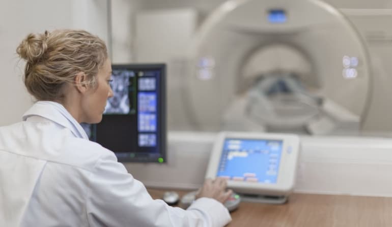
What Is a CT Scan for Children?

A pediatric CT scan is a diagnostic medical imaging procedure that creates three-dimensional images of internal organs, soft tissues, blood vessels, and bones. Compared to the traditional x-ray, it provides a more detailed image that allows medical professionals to diagnose an illness or an injury more accurately. 


Most CT scans take a few seconds, although sometimes the procedure can take more than 10 minutes. 


The medical imaging test uses a large machine that resembles a giant donut with a sliding bed inside where the patient lies still during the procedure. 


The machine emits low-dose radiation that passes through the body and gets picked up by electronic detectors located on the opposite side of the frame to create detailed images. Then, a dedicated computer assembles the “slices” of pictures to come up with three-dimensional images that can show internal injury, tumor, or any abnormality. 


Common Reasons Why Doctors Recommend Pediatric CT Scans

CT scans can help medical professionals diagnose a wide range of health conditions and injuries. In children, these are often performed to identify the causes of severe abdominal pain and evaluate the extent of the injury. 


The medical imaging test is also commonly used to diagnose cancer and monitor patients’ response to treatments. 


Because of the highly detailed images, CT scans are commonly used to evaluate the heart and blood vessels of children and even babies. 


These are the other reasons why doctors recommend pediatric CT scans: 

  • Tumor 
  • Pneumonia and other complications from infections
  • Birth defects
  • Appendicitis
  • Inflammatory diseases 
  • Trauma 
  • trauma to blood vessels or lung
  • Stores in the urinary tract 
  • Seizure and other neurological diseases 
  • Hearing loss
  • Bone-related injuries and defects


In emergencies, CT scan is the go-to medical imaging test since it can quickly detect internal injuries and bleeding, allowing ER doctors to perform life-saving treatments. 


Related article: Managing Claustrophobia and Anxiety During Your CT Scan


Preparing Your Child for a CT Scan 

Each patient will receive a specific set of instructions before undergoing CT scans; hence, you should always ask your doctor for detailed recommendations. 


Meanwhile, the guidelines mentioned below are “general rules” for most, but not all, patients. 

  • Your child may be asked to avoid drinking or eating a few hours before the exam; this is a common requirement for patients who will receive anesthesia or sedatives. 
  • Inform your child’s doctor about his previous tests (including CT scans), allergies, medical condition, and health-related history. 
  • Your child should wear comfortable, loose-fitting clothing (without zippers, metal snaps, and belt buckles) during CT scans. 
  • Before the exam, your child should remove metal objects like eyeglasses, jewelry, retainers, hearing aid, etc. 


Important note: Depending on your child’s age and the area that needs to be scanned, anesthesia may be necessary. In many cases, young children who need to be entirely motionless for a prolonged period may need sedation to ensure high-quality images. 


How to Minimize Radiation Risk

While x-rays and CT scans use very low levels of radiation, it remains ideal to avoid unnecessary exposure. Simply put, these imaging procedures should be only reserved for patients who need them. 


If your child needs a CT scan, you may want to go to children’s hospitals where technicians routinely adjust their machines to compensate for a child’s smaller size. By contrast, general hospitals don’t always change the settings of their scanners. 


In addition, you should tell your doctor if your child has had previous tests, including CT scans. In most cases, repeated CT scans should be avoided unless absolutely necessary. 


Who Interprets the Results? 

Radiologists are medical doctors who can interpret the CT scan results of your child. They will send an official report to your kid’s primary physician. 


CT Scan for Children: What Parents Should Know


For more information about CT scans for children, contact Catalina Imaging at (844) 949-1664. They are the leading mobile CT scanners that can help hospitals and other medical facilities provide high-quality healthcare. 

Reasons Why Your Doctor Might Recommend a CT Scan

Reasons Why Your Doctor Might Recommend a CT Scan | Catalina Imaging

A CT scan is a medical imaging procedure that combines a series of X-rays taken from different angles to create a highly detailed image of the soft tissues, blood vessels, and bones inside your body. 


A CT scan machine is well suited for diagnosing diseases and injuries because it provides detailed images. However, it is particularly useful to ER doctors who need to examine traumatic accident victims quickly. 


Why Would Your Doctor Order a CT Scan? 

With the proliferation of medical imaging machines like MRI, ultrasound, and bone scan, you might wonder why your doctor still recommends a CT scan. 

To better help you understand the benefits of a CT scan, we list some of the most common reasons why your doctor might recommend this medical imaging procedure. 


It is a better option than magnetic resonance imaging or MRI in some situations. 

While MRIs and CT scans are almost similar, there are reasons why MRI is not an ideal procedure. For instance, patients with cochlear implants, implanted pacemakers, and intracranial aneurysm clips can’t have this procedure that involves the use of strong magnets. 

Also, CT scans are shorter and less noisy than MRIs, which emit a banging sound because of the metal coils vibrating during the procedure. 


You have soft tissue damage. 

Compared to traditional X-rays, CT scans provide a crispier, more detailed image of the soft tissue around the bones. And with better images, your doctor can provide a more accurate diagnosis and ultimately a more effective treatment and recovery plan. 


You have a certain type of vascular disease. 

While ultrasound can be used to examine your blood vessels, sometimes their results are inconclusive when it comes to aneurysms, which makes CT scans a better option.

CT scans can also replace surgical biopsy and exploratory surgery, allowing doctors to examine your blood vessels without resorting to surgical procedures. 


You have bone injuries affecting small components. 

If you have bone injuries in your spinal area, hands, feet, and other “small components,” CT scan is a better option than X-rays which may not provide the same level of accuracy. 


You need a bone scan. 

CT scan machines can evaluate your bone density and ultimately determine if you have osteoporosis, which stems from a lifelong lack of calcium. 

CT scans can also diagnose osteoporosis-related fractures that commonly occur in the wrist, spine, and hip. 


You are receiving chemotherapy. 

Cancer patients routinely undergo CT scans to determine if their tumors respond to radiation treatments used to kill cancerous tumors. These machines can also show doctors the tumor’s size and shape. 


You need tumor surgery. 

Before surgery, your specialists may recommend a CT scan alongside biopsy to help them confirm the presence of a tumor and learn about its exact location, shape, and size. The images from the CT scan can also help them examine the tissue surrounding the tumor. 


You just had a traumatic accident. 

While MRIs and CT scans have almost the same functions, CT scans are a better imaging tool in emergencies such as a car collision and slip and fall accident in which doctors need to detect internal injuries quickly to save their patients. 


You need your brain examined. 

Brain CT scans are used to evaluate the presence of mass, tumor, ischemic process (like stroke), fluid collection, hemorrhage, and trauma. Compared to standard X-rays of the head, these machines provide more detailed information about the brain structure and its soft tissue. 

CT scans for the brain usually take between 15 and 30 minutes. 


Related article: Managing Claustrophobia and Anxiety During Your CT Scan


To learn more about CT scans, contact Catalina Imaging at (844) 949-1664 or email them at info@catalinaimaging.com. They provide a mobile CT scan fleet for medical facilities in California, North Carolina, Minnesota, and Illinois

Managing Claustrophobia and Anxiety During Your CT Scan

Managing Claustrophobia and Anxiety During Your CT Scan | anxious child before CT Scan | blog article by Catalina Imaging

Some patients experience signs of claustrophobia during their CT scan, MRI, and or certain types of imaging procedures that require them to lay still in a confined space. Fortunately, there are ways to eliminate or at least minimize their level of anxiety. 


Coping with Scan Anxiety and Managing Claustrophobia

What Is Claustrophobia? 

This type of anxiety disorder results in intense, irrational fear in tight spaces such as elevators, crowded rooms, and medical imaging machines like MRIs and CT scans. Experts suggest that it might stem from traumatic events like experiencing turbulence when flying or being trapped in an enclosed area like cabinets.

Meanwhile, some psychiatrists believe that claustrophobia or any intense, irrational phobia might be related to dysfunction of the amygdala, a part of the brain that controls how you process fear. 


Claustrophobia is Different for Everyone 

As with any type of phobia, claustrophobia is different for anyone in terms of the level of intensity, ranging from mild nervousness to a full-blown panic attack in which the symptoms are almost similar to heart attacks: shortness of breath, numbness of feet and hands, dizziness, fainting, choking sensation, chest pain, trembling, and heart palpitations. 

Some people who have experienced extreme symptoms of claustrophobia describe the feeling as if they’re going to die or the world is going to end. 

Interestingly, many people with claustrophobia know that their intense fear is irrational.

Aside from symptoms and intensity, the triggers also differ from person to person. For instance, some may feel extreme anxiety when riding in an elevator, although plane travel is a “tolerable” experience for them. In contrast, some people can’t travel via aircraft or boat because of their intense phobia, but for some reason, they only experience mild [and manageable] anxiety when using an elevator or being in a small crowded room.


Why Imaging Procedures Might Trigger Claustrophobia 

CT scans, MRIs, and bone scans require enclosed or semi-enclosed medical imaging machines to capture images of the area of concern. Due to the tight space, some patients may experience high levels of anxiety and fear, making it difficult for them to stay still.

It is important to stay perfectly still during medical imaging because even the slightest movement can blur the image. Some machines like CT scans of the abdomen and chest even require patients to hold their breath between 10 and 25 seconds. 

Aside from the tight space, intense anxiety during a medical imaging procedure may also stem from the loudness or “weird” beeping sound of the machine. Some claustrophobic patients have also reported fear of being injured or suffocated during their CT scans or MRIs. 

Some patients have also experienced high anxiety levels after the procedure as they wait for their test results. 


How to Reduce Scan-Related Anxiety 

The list below could provide a better patient experience: 


Mild sedatives 

 If you have a history of anxiety disorder or have claustrophobia, your doctor might recommend a mild sedative to help you stay relaxed during your medical imaging procedure. Sometimes, medications are prescribed alongside a psychotherapy treatment. 

Drugs that may be used right before a medical imaging procedure include beta-blockers, Benzodiazepines, and selective serotonin reuptake inhibitors or SSRIs. 


Soothing music 

If you’re having an MRI scan, you can ask your technician to play a soothing playlist. (Note: MRI machines are particularly loud, which may bother some patients.)


Consider Alternatives 

If you are bothered by the banging noise from MRI machines, you may want to ask your doctor if a CT scan is a good alternative. 

Aside from being shorter and less noisy than MRIs, CT scans are better at providing accurate images that can help your doctor diagnose your injury or disease more easily. 

On the other hand, if your main concern is the tight space, ask your doctor if you can have an ultrasound or an open upright MRI instead. 


Final Thoughts on Anxiety Management During Your CT Scan

Good communications between technologists and patients also play a critical role in reducing anxiety and symptoms of claustrophobia. In general, people who receive specific details (e.g., how long the test will take and how it will be like) will have a better experience than those left in the dark. 

To learn more about better patient experience during a medical imaging procedure, contact Catalina Imaging at (844) 949-1664 or email them at info@catalinaimaging.com. They have mobile CT scans in multiple locations like California, North Carolina, Illinois, and Minnesota.

Medical Imaging for Heart Disease

Medical Imaging for Heart Disease |blog article by Catalina Imaging

In the past, medical imaging to detect heart disease involved expensive and less accurate procedures. Some methods like treadmill stress testing are not recommended or even dangerous for some patients, particularly those with unstable heart failure, symptomatic severe aortic stenosis, and uncontrolled arrhythmia. 


But with the advancement in medical imaging, today’s patients have more options, most of which are safer, more accurate, and even cheaper. 


New imaging technologies that can help doctors diagnose heart disease include the following: 

  • Modern CT Scans
  • PET combined with CT Scan
  • Three-dimensional echocardiography, which is also called 3D echo
  • Magnetic resonance imaging or MRI 


Modern CT Scans

Modern CT scanners can perform a procedure called computed tomography angiography or CTA, which is notable for its accuracy (up to 95%) when detecting small blockages in coronary arteries. The imaging technology also allows doctors to rule out congenital abnormalities and severe heart and artery disease. 


CTA is also significantly less invasive than older techniques like the cardiac cath that involves inserting a long, thin tube called catheter into an artery or vein in the neck, groin, or arm to access the heart. 


In essence, CT scanners are like X-ray machines that provide better imaging. Before the procedure, a contrast dye is injected into the patient’s arm so the scanner, which looks like a giant donut, can take multiple images to create a more detailed impression of the heart and its blood vessels. 


PET Combined with CT Scan

Positron emission tomography or PET scan is an imaging procedure that uses a radioactive substance to detect poor blood flow, buildup of substances in the heart muscle, and other abnormalities in the heart. And when combined with modern CT scanner, the imaging technique can assess not just the heart anatomy but also its function. 


MRI Heart Scan

Compared to earlier medical imaging technologies to detect heart diseases, MRI heart scanners provide more detailed images that can help doctors evaluate the heart structure and function. 


MRI is particularly useful when it comes to showing valve disease, the heart’s overall appearance (volume, shape, and size), tumors, blood clots, and other abnormalities the older imaging technologies may not detect easily. 


While MRI does not use radiation, it relies on powerful magnets to create detailed images. For this reason, the imaging technique is not performed on patients with a defibrillator or pacemaker. 


Nowadays, MRIs come in two versions: open and closed. Open MRI scanners were initially developed to accommodate people with claustrophobia, although they are not an option for medical imaging for heart disease because the organ is in constant motion. 


Echocardiography 

Echocardiography uses high-frequency sound waves called ultrasound to evaluate the valves and heart muscle. Just like in a prenatal ultrasound, it uses a wand-like device that transmits sound waves against the chest area, producing moving images of the heart. 


Because the imaging technology does not use any contrast medium or radiation, it can be performed multiple times without predisposing patients to risk of complications. 


The list below explains the three types of echo test used today:  

  • Three-dimensional echo. This provides multiple ultrasound images of the heart, allowing cardiologists to see a complete image of the heart in motion. Due to its accuracy, it is ideal for evaluating the heart muscle function and its valves. 
  • Portable echo. This inexpensive laptop-sized echo machine is commonly used by paramedics and emergency physicians. 
  • Intracardiac echo. It is used during a cardiac cath procedure in which a long, thin tube is inserted and threaded through the heart to perform surgeries such as closing a hole, opening a narrow valve with a balloon-like device, etc.


Final Word on Imaging for the Heart 

The best imaging procedure for the heart boils down to the symptoms, the patient’s medical condition, and the use of adjunct treatments. To learn more about the topic, contact Catalina Imaging at (844) 949-1664 or info@catalinaimaging.com


Catalina Imaging is one of the leading CT mobile imaging providers in the country, serving hospitals and other healthcare facilities in California, Minnesota, North Carolina, and Illinois. 


How to Prepare for Your CT Scan

How to Prepare for Your CT Scan | patient consulting with doctor | blog article by Catalina Imaging

A CT Scan is a medical imaging procedure used to detect muscle and bone disorders, internal injuries, and certain diseases. Oftentimes, it requires preparation to ensure your comfort and avoid nausea and discomfort while being scanned. 


How to Prepare for Your CT Scan

The preparations before a CT scan may differ from patient to patient, so it is best to always consult with your doctor. In addition, there are instances when a person can’t prepare for an exam; this is particularly true for a car accident victim or anyone who suffers from traumatic injuries who generally needs an immediate imaging scan. 


Food Restrictions

If you are scheduled for a CT scan, your doctor may ask you to avoid drinking and eating at least four hours before your exam. The idea is to eliminate or at least reduce the risk of discomfort and throwing up while lying down.  


Food restriction is particularly important if you’re going to receive an injection of contrast dye, which is used if there is a need for a clearer image of the chest area. For some patients, the solution causes them to feel nauseated, so it is ideal to come to the exam with an empty stomach.  


Blood Test 

Your doctor may recommend a blood test before your scheduled CT scan to ensure that the right contrast dye will be used during the procedure. 


Allergy Medications

Patients allergic to contrast dye injection, which contains iodine, may need to take a steroid medication the night before and morning of the scheduled CT scan. Sometimes, doctors also include antihistamine drugs (e.g., Benadryl) to further eliminate the risk of allergic reactions. 


Oral Solutions

Depending on why you’re getting an exam, you may need to consume a large glass of oral contrast dye, which is a liquid containing Gastrografin or barium. This prep solution is commonly used for patients who need their abdominal area scanned. 


(Note: While most patients are instructed not to eat solid foods, they are generally allowed to drink water, decaffeinated tea or coffee, and juice.)


Arrive Early 

A good rule of thumb is to arrive at least 30 minutes before your CT scan to allow the examination to stay on schedule.


Wear Comfortable, Loose-Fitting Clothing to Your Exam

While your healthcare provider will most likely give you a gown, it still makes sense to wear comfortable clothes to your exam. Also, don’t wear metal objects like eyeglasses, jewelry, hairpins, clothes with metal zippers and buttons, or even dentures while inside the tunnel-like part of the CT scan because they might affect the images. 


Related article: Managing Claustrophobia and Anxiety During Your CT Scan

During the Exam 

During the test, you will lie on your back on a table that slides through a tunnel-like scanner. If your exam requires an IV contrast dye, your healthcare provider will inject it into your arm. 


The contrast dye injection may result in nausea, a “flushed” sensation, or even a slight metallic taste in your mouth. 


Once you’re inside the tunnel-like scanner, you will need to stay as still as possible to avoid blurring the images. On average, the scan itself only takes between 10 and 30 minutes.


After the CT Scan 

You can return to your normal activities after your scan. However, your healthcare provider may ask you to stay for a few minutes to ensure that you don’t feel any discomfort or nausea, which is a common mild side effect of contrast dye. To help your kidney remove the contrast dye from your body, he may also require you to drink lots of water. 


To learn more about mobile CT scan, contact Catalina Imaging at (844) 949-1664. We are a reputable mobile CT company based in Placer County, California, with a combined 50 years of experience. 

CT Scan Rates Continue to Rise due to Patient-Generated Demands

CT Scan Rates Continue to Rise due to Patient-Generated Demands | blog article by Catalina Imaging

Over the past two decades, there has been a notable increase in CT scan rates due to contributing factors such as higher patient-generated demands, improvements in medical imaging technology, and an aging population.


A longitudinal study published in PMC showed that between 1997 and 2006, all medical imaging modalities increased in the US, with CT scan posting one of the highest with 14% yearly growth. This means that over the ten-year period, the number of patients who had the procedure more than doubled. 

CT Scan Rates Continue to Rise due to Patient-Generated Demands


Patient-Generated Demands 

Marketing research company Nielsen conducted a survey suggesting that today’s consumers are “willing to pay premium prices” to achieve their health goals. While millennials are the most proactive age group, the report has also shown that baby boomers–people born between 1946 and 1964–are also more engaged in their quest for “healthy aging” and “active and independent retirement.”  


Today’s more health-conscious consumers, according to some researchers, may be contributing to the increasing demand for CT scan and other medical imaging procedures that promote early diagnosis of diseases.


With early diagnosis of illnesses, California’s leading mobile CT scan provider Catalina Imaging says that “patients can access immediate treatments, which results in high success rate and lower mortality rate.”


In addition, patients are becoming more involved in their healthcare, with many even insisting on a more individualized approach. 


Aging Population 

Surveys have shown a strong correlation between CT scan patients and old age; this is not surprising since aging is a disease risk factor. 


Meanwhile, the US population is aging rapidly due to decreasing childbirth rates and longer life expectancy. In a 2017 US Census Bureau report, by 2030, when every baby boomer will reach the age of 65 years old, one out of five Americans will be of retirement age.  


And by 2035, the report projected that there would be around 78 million people aged 65 years and older versus 76.7 million under the age of 18. This means that within a few years, there will be more seniors than children for the first time in the country’s history. 


The aging population is not the only thing that contributes to the growing need for CT scans. Reports have also shown that the increasing popularity of homecare among the older population is another reason for the growing demand for bedside imaging. 


Improvement in Medical Imaging Technology

While CT scanners have been around since the early 70s, it was only in the late 90s when technological advancements made it possible to “shrink” their size, giving birth to mobile CT scans. 


Portable CT scanners–i.e., they are mounted on wheels and typically run on batteries–allow bedside imaging to become possible and fill in the gap left by fixed CT that is not accessible by some patients living in rural or remote areas. 


Aside from portability, modern CT scan designs produce faster and higher quality images, allowing doctors to diagnose patients with better accuracy and perform treatments with greater precision. 


How Mobile CT Improves Rural Healthcare 

In general, rural hospitals are only seen as critical care providers due to their outdated equipment, medical staff shortage, and other factors contributing to the healthcare disparity between city and rural areas. Furthermore, traveling for care has become a common practice even though this is not a viable option for all patients. 


But with the advent of mobile CT scanners and other similar equipment, rural hospitals can now better serve local patients, especially those in-home care. 


If you are a hospital administrator or radiology manager who needs a mobile CT scan, contact Catalina Imaging at (844) 949-1664 or at info@catalinaimaging.com. Our office is located at 3311 Swetzer Rd, Loomis, CA 95650.

How Long Should You Wait for Your MRI or CT Scan Results?

How Long Should You Wait for Your MRI or CT Scan Results? | blog article by Catalina Imaging

While several factors affect how long you should wait for your MRI or CT scan and the results of these imaging tests, your “priority level” has the most influence. For instance, patients in an emergency situation are not included on the waiting list and can expect to undergo the procedure and receive their results within just a few hours. 


For non-emergencies, the waiting period varies significantly–one day, within a week, or a few months–because radiologists consider some key factors. 

  • Your symptoms and medical history 
  • The complexity of your medical examination (Do you need multiple tests so your doctors can make a comparison?)
  • The transmission between practice to doctor (Even if the radiologists complete the scans within 24 hours, sometimes patients may have to wait longer for their doctors to interpret the results.)


How Long Should You Wait for Your MRI or CT Scan Results?


Turnaround Time: How Long Is Too Long?

Studies have shown that delay in delivering scan results causes the vast majority of patients to experience anxiety, as they interpret it as “bad news.” Thus, leading medical imaging service Catalina Imaging offers Mobile CT units that can travel to hospitals or any convenient sites. 

The company’s Mobile CT Scanners have a spacious changing room and top-notch imaging technologies for quick and accurate scanning. 

Usually, the scans are given to the patients on a disc after the imaging diagnostic exam is complete. However, the turnaround time for their analysis depends on the availability of the radiologists who read the results and the doctors who interpret them. 


In a 2017 report involving around 200 patients, the vast majority wanted their results in one to three days and would call their doctors between one and five days if they had not received news about them. 

For patients who had an MRI or CT scan to check for pneumonia, brain tumor, and cancer treatment, they said they expected results within a day, while those who required it as part of their routine screening and had it to identify the cause of chest pain, they expected results three days and two days, respectively.  


How the Medical Industry Is Dealing with the Backlogs 

With some states still struggling with the increasing number of coronavirus patients, some hospitals have a high volume of results that need to be analyzed and processed. To cope with the backlogs, they prioritize patients who need rapid imaging tests, i.e., those who had been in an accident, had a stroke, or any emergency situation requiring swift medical attention. 


What You Can Do to Reduce Your Waiting Time

While you have no control over some external factors (such as the availability of your provider), there are ways that may help you reduce your wait time. 

  • Opt for a mobile MRI or CT scanner, which is particularly useful when there is a sudden increase in the demand for imaging. 
  • Tell your doctor or the radiology department to call you if there is a last-minute opening on their “cancellation list.”
  • Inform your provider that you are willing to travel to an imaging center with a quicker turnaround time. 
  • Tell your provider that you are willing to go for an appointment in an “ungodly” hour. (Remember, some imaging centers operate 24/7.)
  • Follow your doctor’s food/water intake instructions before your MRI or CT scan to avoid having your test rescheduled. 

The Difference Between CT and MRI

CT vs. MRI: What to Use and Why | blog article by Catalina Imaging

CT vs. MRI: What to Use and Why

While CT and MRI are both medical imaging tools, they use different methods to produce pictures. A CT scan is ideal for larger areas, whereas MRI is better for tissue examination thanks to its more detailed images.


CT vs. MRI: What to Use and Why

How CT Scan Works

CT scans work by taking multiple X-ray images at different angles to create a three-dimensional representation of an internal organ. With a computer, the X-rays are synthesized to form a 3D model of an organ system.

How MRI Scan Works

MRI uses radio waves and magnetic fields to form a detailed image of the bones and soft tissues, providing more thorough imaging than CT scans. 

Which Method Works Best? 

Multiple factors come into play to determine which imaging procedure is ideal and which one should be avoided due to the risk of complications. 

  • Patients who are pregnant should avoid CT scans due to the risk of side effects to their unborn child. 
  • Doctors who need more detailed images recommend MRI. 
  • The type of disease the patient has determines which imaging tool is better. 
  • The medical reason for the imaging determines whether MRI or CT is a better choice. For instance, gunshot and shrapnel victims are advised against having MRI due to the risk of injury, especially if their wound is near or at a sensitive area. 
  • Patients who have claustrophobia may not tolerate the standard MRI scans that use a capsule-shape bed. A possible alternative is the open upright version, although its magnet is not as strong as the traditional MRIs.   
  • Patients with some types of metal implants cannot have MRI scans due to the risk of injury. RF magnetic field can heat up pacemakers and long wires and cause magnetic metals such as intracranial aneurysm clips and some contraceptive devices to react. 

People with cochlear implants, implanted drug infusion pumps, bone-growth stimulators, and some types of prosthetic devices are advised against having MRI scans. However, common dental implants such as Titanium, titanium alloy, and zirconia are safe for MRIs. 

CT and MRI Scans: Detecting Diseases and Medical Conditions 

Both CT and MRI scans can see bones, bone structures, and soft tissues, making them useful in diagnosing a wide range of diseases and medical conditions. But in terms of accuracy, MRI provides more detailed imaging, especially of the soft tissue. 

“Minute” soft tissue injuries like a herniated disc–which CT scans may not be able to detect–are also best diagnosed with MRI scan thanks to its more detailed imaging capabilities. 

Assuming that the patient has no implant or condition that is contraindicated with RF magnetic field, both CT and MRI scans are useful in diagnosing the following diseases and medical conditions: 

  • Head and facial trauma 
  • Ischemic and hemorrhagic stroke
  • Unexplained pain in a certain body area
  • Internal organs such as the bladder, pancreas, stomach, colon, small intestine, uterus, ovary, etc. 
  • Traumatic injuries to the bones 
  • Lumps or tumors
  • Cancer growth 

While CT and MRI scans are both used in cancer diagnosis because they can show tumors or “masses,” they are not a definitive tool. They are mainly used to detect which site is the best to collect a tissue sample for biopsy. 

Once the biopsy has determined that the tumors are cancerous, both imaging techniques remain useful in identifying where the malignant cells have spread or metastasized in the body.

Situations Where CT Scan Is Better 

In emergency situations like stroke, CT scans are a more helpful diagnostic tool than MRIs because they can rapidly scan the brain to identify the underlying cause of stroke (hemorrhagic vs. ischemic). 

MRI scans, meanwhile, are best reserved for non-emergency situations. 

Another issue with MRI is the cost barrier. In general, it costs twice as much as a CT scan, especially if it requires the use of a contrast dye (the average cost of CT is $1,200 versus $2,000 for MRIs). 

CT vs. MRI: What to Use and Why

Final Words 

While MRI and CT scans are great diagnostic imaging tools, MRIs are better in terms of image accuracy and quality. However, people with certain metal implants, gunshot victims, and emergency situations are best diagnosed with CT. 

Canon Medical’s Advancements with AI-based Image Reconstruction Technology

Canon Medical’s Advancements with AI-based Image Reconstruction Technology by Catalina Imaging

New advancements have been made in technology: Canon Medical further utilized Artificial Intelligence to improve image quality that can cater to more patients.

The company announced the further expansion of its Advanced intelligent Clear-IQ Engine (AiCE) Deep Learning Reconstruction (DLR). They used various amounts of high-quality image data to train AiCE, and it can now reduce noise and quickly deliver sharp, clear, and distinct images by boosting its signal. The company’s advancement in this technology features more modalities, clinical indications, and systems.

According to Imaging Technology News Online, this DLR imaging technology has the widest availability, providing advancements in multiple imaging exam types.

AiCE DLR will be showcased for the Cartesion Prime PET/CT system (pending 510(k) clearance) by Canon Medical at the RSNA annual meeting to bring advancements of Artificial Intelligence to molecular imaging. Possible benefits of this include faster scan times, lower dose, and improved image quality.

Canon Medical’s Vantage Orian 1.5T MRI system is now equipped with AiCE DLR for virtually all types of clinical exams that has expanded the capacity to scan clinical indications combined with technologies like Compressed SPEEDER that provides a boost of image quality for clinicians using MR.

Scanning has been expanded to cover a vastly larger number of clinical indications from the prostate to shoulder scans, including all joints, cardiac, pelvis, abdomen, and spine with better image quality. According to Mass Device, Vantage Orian can differentiate true signals from noise using an algorithm, and it can suppress the noise during image reconstruction.

Canon aims to cover the maximum amount of patients that can benefit from AI-based Image Reconstruction Technology. The Senior Vice President and Chief Sales and Marketing Officer of Canon Medical Systems USA, Satrajit Misra, has emphasized the importance of enhanced images for better and easier readability. Therefore, Canon’s efforts to bring accessible AI for technological advancements aim to help clinicians cater to patients better and create a big impact in terms of patient care. Misra also emphasized that the expanded availability of AiCE also aims to expand clinical capabilities as well as seamless integration.

We have good news for you! Catalina Imaging has the latest Canon technology! Let us provide you with better image quality to deliver the best quality service to your patients. We specialize in supplying state-of-the-art mobile CT scanner technology! Learn more about Catalina Imaging and our services. Contact us at (844) 949-1664.

COVID-19 Findings as Collected by Johns Hopkins Experts

COVID-19 Findings as Collected by Johns Hopkins Experts | Catalina Imaging

Cleaning and disinfecting procedures have become more important in several industries since the COVID-19 pandemic happened, particularly in the healthcare sector. Standard procedures and protocols have been revised to better adapt to the coronavirus situation, and scientists are continually looking into new ways to improve these procedures.

Medical experts at John Hopkins have been exploring the use of ultraviolet (UV) light in disinfecting medical equipment, particularly CT scan machines. The inner bore of CT scan machines is more exposed to exhaled particles from the different patients that use them, making them highly susceptible to contracting the virus. While the most common UV rays that hit the earth are classified ultraviolet-A, this light can cause skin cancer and other problems upon exposure, so Siewerdsen and Mahesh opted to test ultraviolet-C (UVC) rays instead since they’ve been known to eliminate a high proportion of SARS-CoV-2 virus from hard surfaces.

Biomedical engineer Jeff Siewerdsen and radiologist Mahadevappa MaheshIt utilized a lamp that emits UVC light to the bed inside the CT scanner to determine if it can successfully reduce virus particles. The experiment proved to be successful, as early results revealed that the UVC light coming from the $105 lamp project was able to eliminate 99.9999% of SARS-CoV-2 virus particles inside the bore in just three to five minutes. They published a summary of their study, accompanied by a video abstract, on November 18 in the Journal of Applied Clinical Medical Physics.

On a different note, researchers have been repeatedly testing for SARS-CoV-2 during the ongoing pandemic in order to isolate the COVID-19 patients and to lower the number of infected individuals. The tests are usually performed on people that are manifesting symptoms or to the ones that were exposed to COVID-19 positive individuals, even without any symptoms.

However, recent research has revealed that even though the genetic material of SARS-CoV-2 can be detected through molecular testing after a few weeks of symptoms, it does not mean that infectious virus particles are present. A study by researchers at Johns Hopkins Medicine and the Johns Hopkins Bloomberg School of Public Health has provided more understanding about the detection of the virus while also showing its contagiousness.

In their study, the researchers evaluated almost 30,000 nasopharyngeal swab results of repeated polymerase chain reaction (PCR) diagnostic tests. The number of times a PCR takes to get a positive signal is called the cycle threshold (Ct). A low Ct score indicates a large amount of SARS-CoV-2 RNA and a high one means a fewer amount of the genetic material.

The experiments’ results revealed that the average Ct value associated with cell culture growth of SARS-CoV-2 was 18.8. The researchers also detected viral growth from specimens collected up to 20 days after the first positive result. Sequencing of the entire genome from RNAs collected in the first and subsequent tests provided evidence that the same virus was seen throughout. Positive tests following negative ones had Ct values higher than 29.5, and were not associated with observed virus growth in culture.

According to Dr. Andrew Pekosz, professor of microbiology and immunology at the Johns Hopkins Bloomberg School of Public Health and co-senior author of the study, additional studies need to be conducted to confirm if Ct values and cell cultures are accurate enough to make clinical decisions for diagnosis. “Defining the window of time in which a COVID-19 patient can transmit the virus can help drive more effective isolation practices,” Dr. Pekosz explains.

(Source: Science Codex)