Computed Tomography (CT) scans—or computerized axial tomography (CAT)—have become an essential part of modern healthcare due to their precision and adaptability. These diagnostic tools have been used in medical imaging for several decades and have undergone improvements to increase safety and reduce radiation exposure since the 1970s.
CT scans enable healthcare professionals to detect and diagnose various medical conditions with unmatched accuracy.
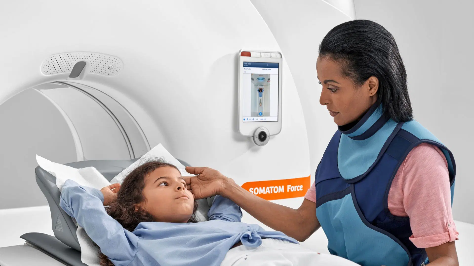
And what makes CT scans even more remarkable is their portability, thanks to the advent of mobile CT scanners, which made these advanced imaging capabilities accessible at the bedside, in emergency rooms, and even in remote locations.
Mobile CT scanners are transforming the way medical imaging is conducted, saving lives, improving patient care, and enhancing healthcare efficiency. In this article, we’ll explore the history and mechanisms behind CT scanners and how they’re empowering healthcare providers to deliver exceptional care.
Harness the potential of mobile CT scanners for your healthcare needs! Our top-tier mobile CT rentals at Catalina Imaging can empower your medical facility with advanced imaging solutions. Contact us at [email protected] or (844) 949-1664 to learn how we can assist you in providing the best care for your patients.
Although CT scans are associated with science and technology, their origins may surprise you. The pioneering technology has a fascinating connection to rock and roll, specifically the phenomenal success of The Beatles in the 1960s.
Rumor has it that Electric and Music Industries (EMI), which owned Abbey Road Studios and catapulted the band to stardom, channeled the enormous profits from The Beatles’ albums in the 1960s into funding pioneering research. At their peak, The Beatles’ record and ticket sales earned approximately $650 a second in today’s money.
But EMI was more than just a record label; they had a significant presence in the electrical industry. In 1959, they launched the EMIDEC 1100, a commercial computer that marked their foray into technology. They also invested in medical equipment research, which eventually led to a groundbreaking innovation.
Godfrey Hounsfield, a key figure in the EMIDEC project, began developing the first medical scanner. With substantial support from the UK government (£600,000, equivalent to £7 million today), Hounsfield and his team spent four years inventing and building the first computed tomography scanner.

In a remarkable twist, fans who grooved to hits like “Can’t Buy Me Love,” “PS I Love You,” “Love Me Do,” and their iconic cover of “Twist and Shout” inadvertently contributed to this revolutionary technology.
In 1972, British engineer Godfrey Hounsfield co-invented the technology with physicist Dr. Allan Cormack. They were jointly awarded the 1979 Nobel Prize in Physiology and Medicine. Hounsfield was knighted in 1981, becoming Sir Godfrey Hounsfield.
However, the mathematical foundations of the CT scanner were laid by Johann Radon in 1917 with the “Radon transform” and Stefan Kaczmarz’s “Algebraic Reconstruction Technique” in 1937. Hounsfield built upon these theories to create a groundbreaking medical advancement.

Despite his remarkable achievements, Hounsfield had no formal qualifications, having left school at 16. Hence, all the degrees bestowed upon him were honorary. He also never got married, claiming not to have established a permanent residence until he was 60. Initially, his work at EMI focused on radar and guided weaponry, and his peers described him as a “crank.” Hounsfield passed away in 2004 at 84.
Hounsfield’s idea for the CT scanner came to him while on vacation, when he wanted to reconstruct a 3D picture of a box by imagining it as a series of slices. This inspired thought led to further EMI research and funding, and the first commercially viable CT scanner was installed at Atkinson’s Morley Hospital in 1971.
To transition the brain scanner to mainstream medicine, Hounsfield collaborated with consultant radiologist James Ambrose. They created a prototype to study preserved human and animal organs.
The first human patient, a middle-aged woman believed to have a brain tumor, was scanned on October 1, 1971, by James Ambrose. This procedure took several days to complete, requiring meticulous effort and patience.
Each scan, or “slice,” took 30 minutes to capture, and the raw data was stored on magnetic tapes. These tapes were then transported across town, where the data was processed on an EMI mainframe computer over 2.5 hours. Finally, a Polaroid camera was used to capture the reconstructed image, which was rushed back to the hospital.
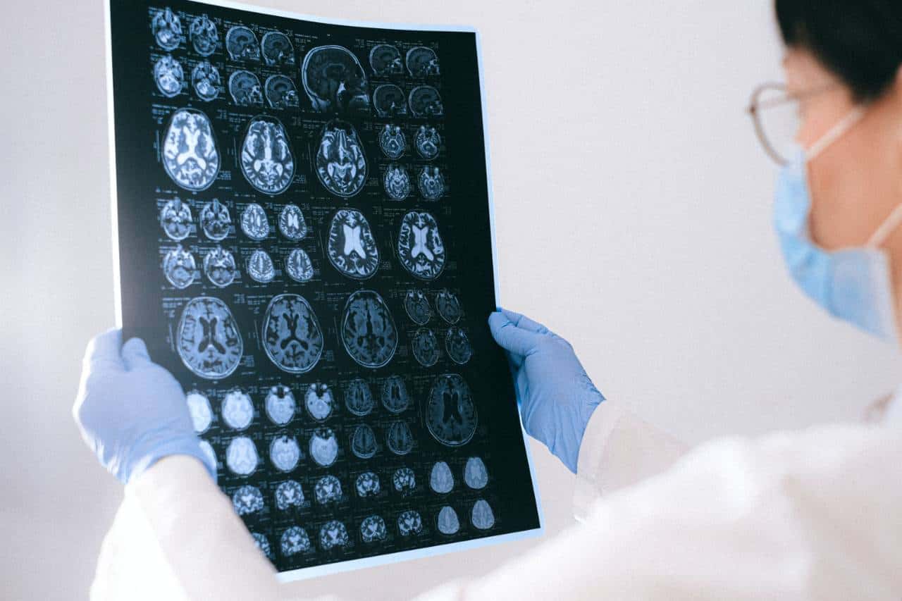
The wait was worth it – the scan revealed a cystic mass, approximately the size of a plum, in the woman’s left frontal lobe. This groundbreaking moment marked the obsolescence of all other brain imaging methods.
The success of the prototype brain scanner at Atkinson Morley Hospital made headlines in 1972, paving the way for a medical revolution. By 1973, the United States had embraced this innovative technology, installing its first CT scanners.
The popularity of CT scans skyrocketed, with a staggering 3 million examinations conducted by 1980, transforming the field of medical imaging forever.

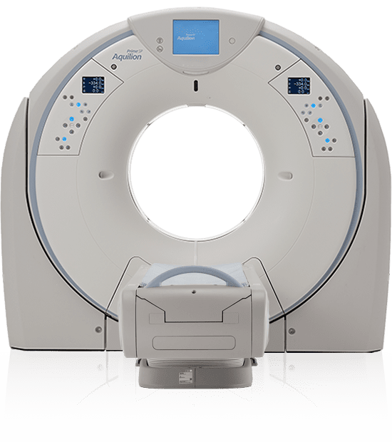
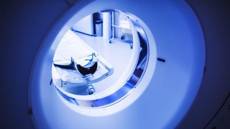
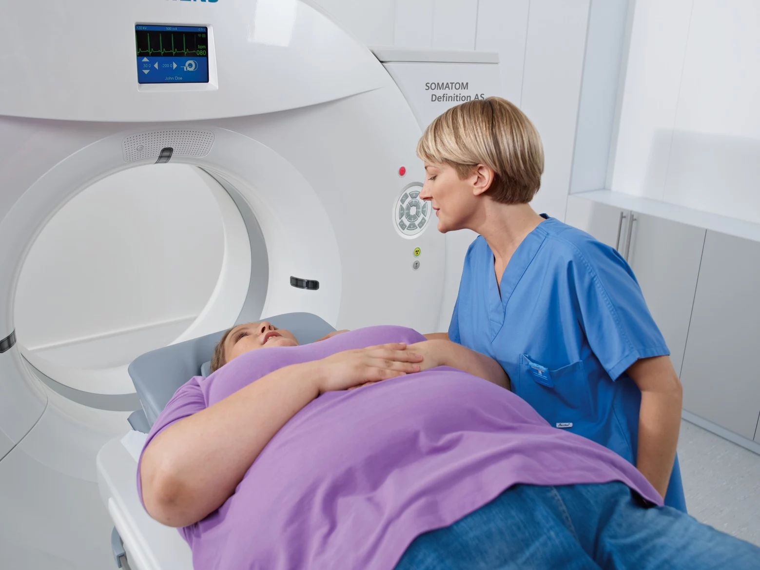
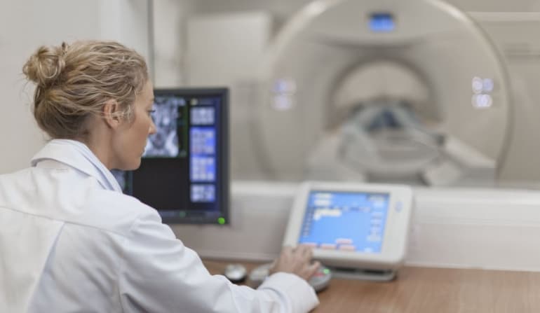
CT scanners work by taking a series of X-ray images from different angles around the body, which are then processed by a computer to create detailed cross-sectional images or “slices.” These slices provide a three-dimensional view of the area of interest, allowing healthcare professionals to examine it from various angles and depths.
The core components and mechanisms that make CT scans possible include:
CT scans are an indispensable diagnostic tool in various clinical settings, offering exceptional versatility in detecting a wide range of conditions and anatomical structures.

Some of the key areas where CT scans are used include:
The remarkable imaging capabilities of CT scans have pushed the medical industry to staggering heights, but a significant gap remains in providing timely access to CT scans in critical situations.
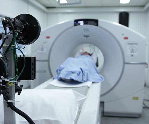
Conventional CT scanners, weighing around 4,000 kilograms and requiring high-voltage power and climate-controlled rooms, have limited their use in battlefields, ambulances, and emergency rooms.
The unmet need for convenient and rapid CT scans, particularly in cases of stroke and brain trauma, has become a pressing concern in the medical community. Despite advancements in imaging techniques, the immobility of traditional CT scanners has been a long-standing pain point.
So, the introduction of portable CT scanners has revolutionized the field.
In the 1970s, Medical Coaches Inc., founded by Ian Smith, launched the world’s first mobile CT scanner in Peru, marking a significant milestone in medical history.
The pioneering mobile CT scanner primarily focused on head scans and attempted to provide cross-sectional images of the heart. Additionally, the mobile healthcare unit offered ultrasound services for pregnant women to assess the health of their hearts, gallbladders, breasts, and livers.
This comprehensive approach to mobile healthcare set the stage for future advancements in portable CT scanners and their impact on medical care.

The demand for mobile CT scanners has skyrocketed in recent years, driven by the urgent need to provide swift care to stroke patients.
A series of incidents highlighted the critical importance of rapid treatment, leading to the exploration of mobile stroke units (MSUs) in 2003.
The concept of “bringing the hospital to the patient” significantly reduced the waiting period from the initial distress call to therapy. More importantly, the presence of a portable CT scanner in these MSUs enabled professionals to treat patients quickly and accurately during emergencies.

In 2010, the University Hospital of the Saarland shared their initial results of the first MSU. They showed that it worked with an average call-to-decision time of 35 minutes.
Many more MSUs have been created and deployed since then. The first MSU in the United States was developed in Houston, Texas, and has been in clinical use since May 2014.
The University of Tennessee Health Science Center has also embraced MSUs and portable CT scanners, equipping their 14-ton ambulance with tools for early fluid infusion and dye blood vessel analysis. Given Tennessee’s status as a “stroke belt,” this technology has been a game-changer for many patients.
The Memphis MSU staff achieves remarkable response times, providing treatment in just 13-14 minutes – a significant improvement over the 40-50 minutes typically spent in emergency rooms.
Neurologists are eager for more portable CT scanners, particularly in intensive care units. The availability of a mobile CT scanner on hand would exponentially increase efficiency, eliminating the need for patient and personnel transfer to fixed CT scanners.
Discover the cutting-edge mobile CT scanners from Catalina Imaging, designed to deliver exceptional image quality and unparalleled patient care. Our state-of-the-art technology ensures precise diagnoses and effective treatment plans. Reach out to us at [email protected] or (844) 949-1664 to learn more about our fleets.
Mobile CT scanners operate on the same fundamental principles as traditional stationary CT scanners but boast a compact and portable design, enabling effortless transport to various locations, such as emergency rooms, operating rooms, or even ambulances.
The mobility of these devices has made it possible to bring CT scanning capabilities to the point of care, a remarkable feat in global healthcare delivery, especially in time-sensitive and critical situations.
Here’s an overview of how CT scans are performed using mobile CT scanners:
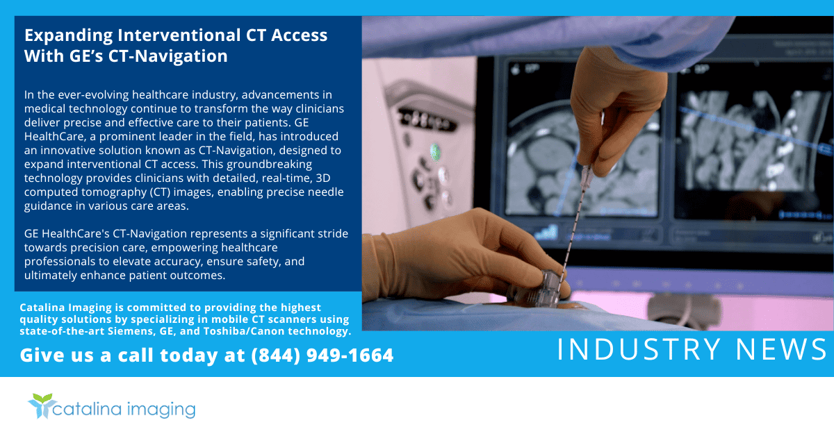
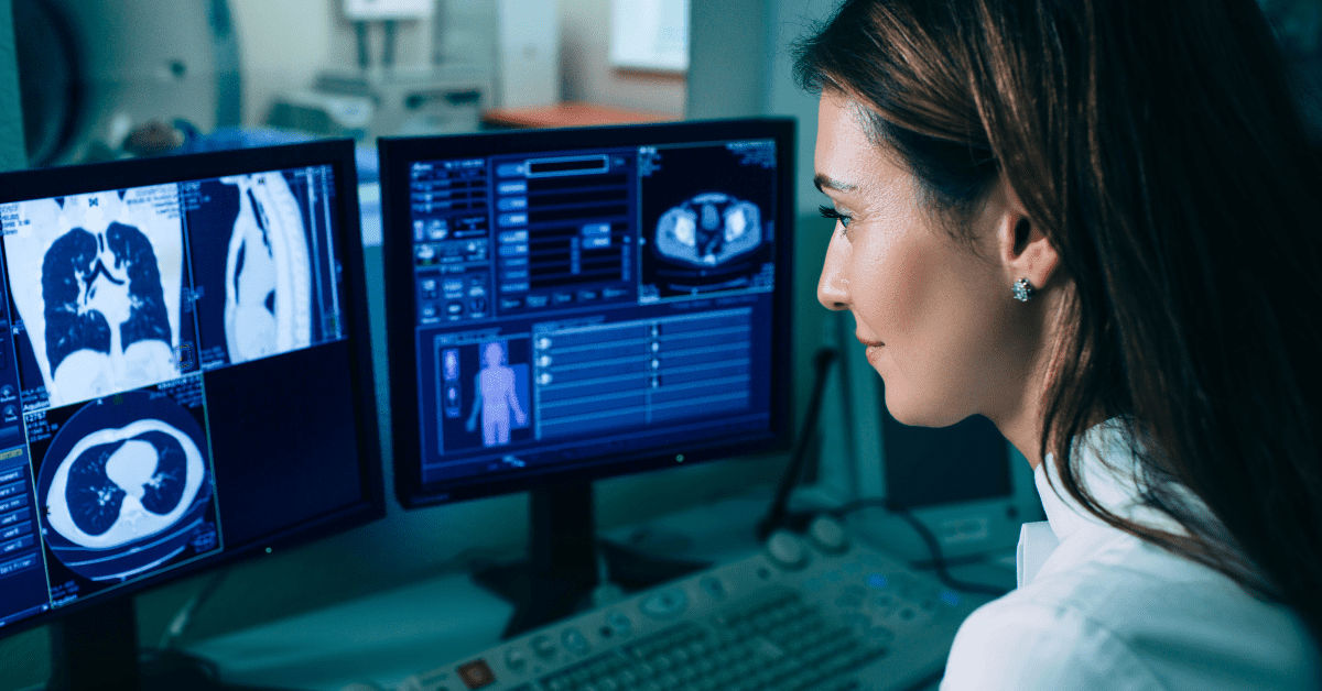
The rapid advancement of Artificial Intelligence (AI) is poised to transform the medical imaging industry, with radiologists and pathologists worldwide set to benefit significantly.

A recent study demonstrates the immense potential of AI in medical imaging, showcasing its capability to detect acute neurologic events in CT scan images in a mere 1.2 seconds.
The remarkable achievement was made possible by analyzing 37,000 head CT exams, with the AI system outperforming human radiologists in diagnosing and identifying neurological conditions like stroke.
This development is critical to improving patient care, particularly in alerting physicians to urgent concerns. As the process is 150 times faster, hours could turn into mere seconds— an advancement that would undoubtedly unburden the hospital staff.
Although the research is ongoing and the AI platform requires real-world testing, this study exemplifies the transformative power of AI in radiology. As AI continues to evolve, it is likely to augment the capabilities of radiologists, enhancing the accuracy and speed of diagnoses and ultimately improving patient outcomes.
Computed Tomography (CT) scans have transformed the medical landscape, offering a wide range of benefits for patients with internal injuries, trauma, and various diseases. This technology enables doctors to visualize almost every part of the body, facilitating accurate diagnoses.
Moreover, CT scans play a vital role in guiding treatment plans, whether surgical, medical, or radiation-based and help doctors monitor the effectiveness of medications and other treatments.
Since its introduction by Hounsfield, CT scan technology has undergone remarkable advancements, becoming an indispensable tool in modern medicine. While we may not yet have the futuristic, instant-diagnosis capabilities of Star Trek, the rapid progress in radiology is bringing us closer to that reality.
Mobile CT scanners, in particular, embody innovation and progress in medical imaging. The ability to bring this diagnostic power directly to the patient’s side has revolutionized emergency and critical care.
As technology continues to evolve, we can expect further enhancements in mobile CT scanner design, ultimately ensuring that everyone, regardless of location, receives the best possible care.
Upgrade your healthcare facility with advanced mobile CT scanners. Catalina Imaging provides top-notch rental services for mobile CT scanners tailored to meet the changing needs of modern medicine. Start your journey towards better healthcare with a simple click or call – reach out to [email protected] or (844) 949-1664 today!